. 2021 Oct 1;77(Pt 10):1317-1335.
doi: 10.1107/S2059798321008937. Epub 2021 Sep 23.

Structural and catalytic characterization of Blastochloris viridis and Pseudomonas aeruginosa homospermidine synthases supports the essential role of cation-π interaction

Affiliations

Structural and catalytic characterization of Blastochloris viridis and Pseudomonas aeruginosa homospermidine synthases supports the essential role of cation-π interaction

F Helfrich et al. Acta Crystallogr D Struct Biol. .

Abstract

Polyamines influence medically relevant processes in the opportunistic pathogen Pseudomonas aeruginosa, including virulence, biofilm formation and susceptibility to antibiotics. Although homospermidine synthase (HSS) is part of the polyamine metabolism in various strains of P. aeruginosa, neither its role nor its structure has been examined so far. The reaction mechanism of the nicotinamide adenine dinucleotide (NAD+)-dependent bacterial HSS has previously been characterized based on crystal structures of Blastochloris viridis HSS (BvHSS). This study presents the crystal structure of P. aeruginosa HSS (PaHSS) in complex with its substrate putrescine. A high structural similarity between PaHSS and BvHSS with conservation of the catalytically relevant residues is demonstrated, qualifying BvHSS as a model for mechanistic studies of PaHSS. Following this strategy, crystal structures of single-residue variants of BvHSS are presented together with activity assays of PaHSS, BvHSS and BvHSS variants. For efficient homospermidine production, acidic residues are required at the entrance to the binding pocket (`ionic slide') and near the active site (`inner amino site') to attract and bind the substrate putrescine via salt bridges. The tryptophan residue at the active site stabilizes cationic reaction components by cation-π interaction, as inferred from the interaction geometry between putrescine and the indole ring plane. Exchange of this tryptophan for other amino acids suggests a distinct catalytic requirement for an aromatic interaction partner with a highly negative electrostatic potential. These findings substantiate the structural and mechanistic knowledge on bacterial HSS, a potential target for antibiotic design.

Keywords: Blastochloris viridis; NAD; Pseudomonas aeruginosa; Rossmann fold; cation–π interactions; homospermidine synthases; polyamine metabolism; putrescine; transferases.


Figures

Figure 1
Figure 1
The main reaction catalyzed by HSS. Two putrescine (PUT) molecules are converted into one sym-homospermidine (HSP) molecule.
Figure 2
Figure 2
Proposed reaction steps in the conversion of PUT to HSP by bacterial HSS. The relevant residues, NAD(H), PUT, HSP and intermediates are shown as two-dimensional structure representations. Hydrogen bonds are depicted as blue dotted lines, delocalized electrons as dashed lines, cation–π interactions as orange dashed–dotted lines and electron transfers as red arrows. Atom numbering for PUT and HSP is given in green. For simplicity, steps 5 and 6 are shown as a combined depiction with correspondingly labeled electron transfers. A more detailed sequence of the reaction steps has been described previously (Krossa et al., 2016 ▸) and additional intervening reaction steps are proposed in Supplementary Fig. S2.
Figure 3
Figure 3
Structure and binding pocket of PaHSS. (a) Cartoon representation of PaHSS (PDB entry 6y87 chain A) with the NAD(P)-binding Rossmann-like domain colored yellow (residues 1–155 and 389–419) and the HSS-like domain in blue (residues 156–388 and 420–469). The solvent-accessible surface of the binding pocket is depicted in transparent red with the entrance pointing upwards. The NAD+ molecule lining the surface of the pocket is shown in ball-and-stick representation. (b) The solvent-accessible surface of the binding pocket was rotated by an angle of ∼90° around the y axis in relation to (a). Selected side chains lining the pocket surface, the NAD+ molecule and the PUT molecule within the active site are depicted in ball-and-stick representation.
Figure 4
Figure 4
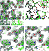
Active sites of three PaHSS molecules with bound PUT molecules. Atomic models of PaHSS molecules [PDB entry 6y87 chain A (a), chain C (b) and chain E (c)] are depicted in ball-and-stick representation with electron-density maps as mesh. The large images show the 2mF oDF c maps (black, 1σ) and mF oDF c maps (green/red, ±4σ) of the complete models. Models lacking the PUT molecules were used to calculate (I) 2mF oDF c composite omit maps including simulated annealing (black, 1σ), (II) mF oDF c composite omit maps including simulated annealing (green/red, ±3σ) and (III) mF o − DF c polder maps (green/red, ±5σ).
Figure 5
Figure 5
Potential NAD adduct in the active site of PaHSS molecule B. The atomic model of the PaHSS molecule (PDB entry 6y87 chain B) is depicted in ball-and-stick representation in two orientations in (a) and (b). The 2mF oDF c map (black, 1σ) and the mF oDF c map (green/red, ±4σ) are shown as mesh. The central, uninterpreted electron-density distribution could originate from a substitution at the nicotinamide ring.
Figure 6
Figure 6
Structural comparison of PaHSS and BvHSS. Superimposition of PaHSS (PDB entry 6y87 chain A, NAD(P)-binding Rossmann-like domain in yellow, HSS-like domain in blue) onto BvHSS (PDB entry 4tvb chain B, gray; Krossa et al., 2016 ▸). Protein models are depicted in cartoon representation with selected side chains and NAD+ molecules in ball-and-stick representation. (a, b) Depiction of the solvent-accessible binding pocket surfaces of PaHSS (red, transparent) and BvHSS (gray, opaque). In (b), the binding-pocket surfaces were clipped and the NAD+ molecules were hidden for clarity. (c, d) Depiction of selected side chains (numbered according to the PaHSS sequence) in the superimposed binding pockets. In (d), the NAD+ molecules are hidden for clarity.
Figure 7
Figure 7
Geometry of cation–π interaction between PUT and tryptophan in PaHSS and BvHSS. Interaction geometry between PUT atoms C4 and N2 and the tryptophan benzene moiety in (a, b) PaHSS (PDB entry 6y87 chain E) and (c, d) BvHSS (PDB entry 4tvb chain B; Krossa et al., 2016 ▸). Structures are shown in ball-and-stick representation, with distances as yellow dashed lines, angle legs as gray lines and angles as gray transparent triangles [not visible for φ in (c)]. The orthogonal projections of C4 and N2 onto the ring planes are shown as black spheres (C4′ and N2′). All angle legs originate from the centroid of the benzene moiety, including the dashed distance vectors (centroid to C4 and N2). The angle θ is spanned by the normal of the ring plane (gray, infinitely pointing upwards) and the C4 or N2 distance vector (yellow, dashed). The angle φ is between the vector pointing to C4′ or N2′ and the vector pointing to the CH2 ring carbon.
Figure 8
Figure 8
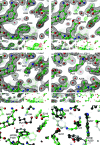
BvHSS variants targeting the ‘ionic slide’ and the ‘inner amino site’. (a) BvHSS variant E117Q (PDB entry 6s6g chain B), (b) superimposition of wild-type BvHSS (PDB entry 4tvb chain B, gray) onto (a), (c) the E210Q variant (PDB entry 6s3x chain A) and (d) the E210A variant (PDB entry 6s49 chain A). Atomic models are depicted in ball-and-stick representation and electron-density maps as mesh. The large images (a, c, d) show the 2mF oDF c maps (black, 1σ) and mF oDF c maps (green/red, ±4σ) of the complete models. Models lacking the respective exchanged residue together with ten N-­terminally and ten C-terminally adjacent residues were used to calculate (I) 2mF oDF c omit maps (black, 1σ) and (II) mF oDF c omit maps (green/red, ±4σ) after refinement including simulated annealing.
Figure 9
Figure 9
BvHSS variants targeting the tryptophan at the active site. (a) BvHSS variant W229F (PDB entry 6s4d chain A), (b) BvHSS variant W229E (PDB entry 6sep chain B), (c, d) BvHSS variant W229A (PDB entry 6s72 chain A) from different perspectives. (e, f) Superimposition of wild-type BvHSS (PDB entry 4tvb chain B, gray; Krossa et al., 2016 ▸) onto BvHSS variant W229F (colored) (e) and BvHSS variant W229A (colored) (f). Atomic models are depicted in ball-and-stick representation and electron-density maps as mesh. Large images (ad) show the 2mF oDF c maps (black, 1σ) and mF oDF c maps (green/red, ±4σ) of the complete models. Models lacking the respective exchanged residue together with ten N-terminally and ten C-terminally adjacent residues [I and II in (ac)] or lacking the PUT molecule [I and II in (d)] were used to calculate (I) 2mF oDF c omit maps (black, 1σ) and (II) mF oDF c omit maps (green/red, ±4σ) after refinement including simulated annealing.

Similar articles

Cited by

References

    1. Anantharajah, A., Mingeot-Leclercq, M.-P. & Van Bambeke, F. (2016). Trends Pharmacol. Sci. 37, 734–749. - PubMed
    1. Becker, N. N. & Roberts, J. D. (1984). Biochemistry, 23, 3336–3340. - PubMed
    1. Benach, J., Atrian, S., Gonzàlez-Duarte, R. & Ladenstein, R. (1999). J. Mol. Biol. 289, 335–355. - PubMed
    1. Bitonti, A. J., McCann, P. P. & Sjoerdsma, A. (1982). Biochem. J. 208, 435–441. - PMC - PubMed
    1. Böttcher, F., Ober, D. & Hartmann, T. (1994). Can. J. Chem. 72, 80–85.

MeSH terms

Supplementary concepts