. 2021 Oct 1;77(Pt 10):334-340.
doi: 10.1107/S2053230X21009195. Epub 2021 Sep 21.

Structural characterization of human peptidyl-arginine deiminase type III by X-ray crystallography

Affiliations

Structural characterization of human peptidyl-arginine deiminase type III by X-ray crystallography

Othman Rechiche et al. Acta Crystallogr F Struct Biol Commun. .

Abstract

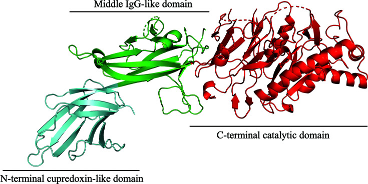
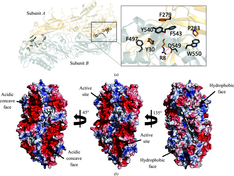
The Ca2+-dependent enzyme peptidyl-arginine deiminase type III (PAD3) catalyses the deimination of arginine residues to form citrulline residues in proteins such as keratin, filaggrin and trichohyalin. This is an important post-translation modification that is required for normal hair and skin formation in follicles and keratocytes. The structure of apo human PAD3 was determined by X-ray crystallography to a resolution of 2.8 Å. The structure of PAD3 revealed a similar overall architecture to other PAD isoforms: the N-terminal and middle domains of PAD3 show sequence and structural variety, whereas the sequence and structure of the C-terminal catalytic domain is highly conserved. Structural analysis indicates that PAD3 is a dimer in solution, as is also the case for the PAD2 and PAD4 isoforms but not the PAD1 isoform.

Keywords: calcium binding; hair follicles; peptidyl-arginine deiminase; post-translational modifications; protein citrullination.


Figures

Figure 1
Figure 1
Cartoon representation of the PAD3 monomer structure. The N-terminal cupredoxin-like domain, the central IgG-like domain and the C-terminal catalytic domain are coloured cyan, green and red, respectively.
Figure 2
Figure 2
The PAD3 dimer. (a) Cartoon representation of two PAD3 molecules arranged in a head-to-tail manner. Residues involved in the head-to-tail dimer interface are represented as sticks. Residues Arg8/Asp549 form a salt bridge and Phe279/Tyr540/Phe543, Phe497/Tyr30 and Pro283/Trp550 form hydrophobic clusters. (b) The whole dimer is shown with a solvent-accessible surface coloured by electrostatic potential, showing the two faces of the molecule. Electrostatic potentials were calculated using APBS (Baker et al., 2001; Jurrus et al., 2018; red =−90 k B T/e c, blue = +90 k B T/e c).
Figure 3
Figure 3
The Ca2+-binding sites of PADs. (a) Ca2+-binding sites and their respective coordinating residues determined from X-ray crystallographic structures of PAD1 (PDB entry 5hp5), PAD2 (PDB entries 4n2b and 4n2c) and PAD4 (PDB entries 1wd8 and 1wd9). A structural alignment of PADs (PAD1, PAD2, PAD3 and PAD4) was conducted to determine the residues in PAD3 that are likely to be involved in Ca2+ coordination. The Ca2+-coordinating residues in PAD1, PAD2 and PAD4 that were visualized by X-ray crystallography (Arita et al., 2004; Slade et al., 2015; Saijo et al., 2016 ▸) are listed in black. The putative Ca2+-coordinating residues in PAD3 are listed in blue. The residues shown to not coordinate Ca2+ in PAD1 and PAD4 are listed in green. (b) Structure superposition of PAD3 (PDB entry 6ce1, light orange) with Ca2+-bound PAD2 (PDB entry 4n2b, purple) and Ca2+-bound PAD4 (PDB entry 1wd9, blue). All Ca2+ sites are shown in red for descriptive purposes. (c) Structure superposition of PAD3 (PDB entry 6ce1, light orange) with apo PAD2 (PDB entry 4n20, hot pink) and apo PAD4 (PDB entry 1wd8, light blue).

Similar articles

Cited by

References

    1. Afonine, P. V., Grosse-Kunstleve, R. W., Echols, N., Headd, J. J., Moriarty, N. W., Mustyakimov, M., Terwilliger, T. C., Urzhumtsev, A., Zwart, P. H. & Adams, P. D. (2012). Acta Cryst. D68, 352–367. - PMC - PubMed
    1. Arita, K., Hashimoto, H., Shimizu, T., Nakashima, K., Yamada, M. & Sato, M. (2004). Nat. Struct. Mol. Biol. 11, 777–783. - PubMed
    1. Arita, K., Shimizu, T., Hashimoto, H., Hidaka, Y., Yamada, M. & Sato, M. (2006). Proc. Natl Acad. Sci. USA, 103, 5291–5296.6. - PMC - PubMed
    1. Assohou-Luty, C., Raijmakers, R., Benckhuijsen, W. E., Stammen-Vogelzangs, J., de Ru, A., van Veelen, P. A., Franken, K. L., Drijfhout, J. W. & Pruijn, G. J. (2014). Biochim. Biophys. Acta, 1844, 829–836. - PubMed
    1. Baker, N. A., Sept, D., Joseph, S., Holst, M. J. & McCammon, J. A. (2001). Proc. Natl Acad. Sci. USA, 98, 10037–10041. - PMC - PubMed

Substances