. 2021 Oct 1;77(Pt 10):348-355.
doi: 10.1107/S2053230X21009523. Epub 2021 Sep 27.

Crystal structures of human coronavirus NL63 main protease at different pH values

Affiliations

Crystal structures of human coronavirus NL63 main protease at different pH values

Hongxia Gao et al. Acta Crystallogr F Struct Biol Commun. .

Abstract

Human coronavirus NL63 (HCoV-NL63), which belongs to the genus Alphacoronavirus, mainly infects children and the immunocompromized and is responsible for a series of clinical manifestations, including cough, fever, rhinorrhoea, bronchiolitis and croup. HCoV-NL63, which was first isolated from a seven-month-old child in 2004, has led to infections worldwide and accounts for 10% of all respiratory illnesses caused by etiological agents. However, effective antivirals against HCoV-NL63 infection are currently unavailable. The HCoV-NL63 main protease (Mpro), also called 3C-like protease (3CLpro), plays a vital role in mediating viral replication and transcription by catalyzing the cleavage of replicase polyproteins (pp1a and pp1ab) into functional subunits. Moreover, Mpro is highly conserved among all coronaviruses, thus making it a prominent drug target for antiviral therapy. Here, four crystal structures of HCoV-NL63 Mpro in the apo form at different pH values are reported at resolutions of up to 1.78 Å. Comparison with Mpro from other human betacoronaviruses such as SARS-CoV-2 and SARS-CoV reveals common and distinct structural features in different genera and extends knowledge of the diversity, function and evolution of coronaviruses.

Keywords: apo structure; coronaviruses; crystallization; human coronavirus NL63; main protease.


Figures
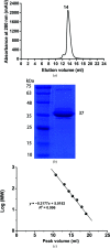
Figure 1
Figure 1
Purification of recombinant HCoV-NL63 Mpro. (a) Size-exclusion chromatography of HCoV-NL63 Mpro. HCov-NL63 Mpro eluted as a single peak from a Superdex 200 Increase 10/300 GL column. The peak volume is about 14 ml. The calculated molecular weight is approximately 74 kDa using the standard curve in (c), suggesting a dimeric form. (b) SDS–PAGE analysis of purified HCoV-NL63 Mpro. The monomer runs as a single band and the purity is greater than 95%. According to the protein standard markers, the molecular mass is close to 37 kDa. (c) A standard curve for SDS–PAGE (a) was generated using known protein standards. MW represents the molecular weight of the corresponding protein. x represents the peak volume. MW (kDa) = 10(−0.2177x + 5.0182).
Figure 2
Figure 2
Crystals of HCoV-NL63 Mpro at four different pH values. (a) 0.1 M sodium citrate tribasic dihydrate, 2%(v/v) Tacsimate, 16%(w/v) polyethylene glycol 3350, pH 5.6. (b) 0.2 M potassion formate, 20%(w/v) polyethylene glycol 3350, pH 7.5. (c) 0.1 M sodium citrate tribasic dihydrate, 16%(w/v) polyethylene glycol 3350, pH 5.0. (d) 0.1 M sodium citrate tribasic dihydrate, 16%(w/v) polyethylene glycol 3350, pH 5.2.
Figure 3
Figure 3
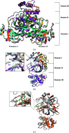
Structure of HCoV-NL63 Mpro. (a) Overview of homodimers shown as cartoons. The coloring is as follows: pH 5.0, orange; pH 7.5, cyan; pH 5.2, magenta; pH 5.6, yellow. (b) Structural alignment of protomer A in Mpro (pH 5.0, cyan; pH 7.5, magenta; pH 5.2, yellow; pH 5.6, orange) with that in the previous reported apo enzyme (gray; PDB entry 3tlo). The backbone is presented in cartoon representation. (c) Comparison of protomer A in structures of apo-form HCoV-NL63 Mpro (orange, PDB entry 7e6r) and HCoV-NL63 Mpro bound to the inhibitor N3 (gray; PDB entry 5gwy). N3 is shown as green sticks.
Figure 4
Figure 4
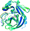
Superimposition of HCoV-NL63 Mpro with Mpro from SARS-CoV and SARS-CoV-2. The main chains of the three Mpro structures (HCoV-NL63, cyan, PDB entry 7e6l; SARS-CoV, green, PDB entry 1uj1; SARS-CoV-2, blue, PDB entry 7c2q) are superimposed and the backbones are represented as cartoons. Residues 45–51 and residues 186–191 are marked with a dashed yellow oval and a red oval, respectively.
Figure 5
Figure 5
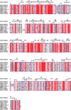
Alignment of the Mpro amino-acid sequences from seven CoVs. The sequence and secondary structure of HCoV-NL63 Mpro (PDB entry 7e6m) are used as a reference for the alignment. ClustalW was used for sequence alignment and ESPript 3.0 was used to generate the graphical representation. Secondary-structural elements shown include α-helices, β-strands and β-turns (TT).

References

    1. Abdul-Rasool, S. & Fielding, B. C. (2010). Open Virol. J. 4, 76–84. - PMC - PubMed
    1. Anand, K., Ziebuhr, J., Wadhwani, P., Mesters, J. R. & Hilgenfeld, R. (2003). Science, 300, 1763–1767. - PubMed
    1. Annan, A., Ebach, F., Corman, V. M., Krumkamp, R., Adu-Sarkodie, Y., Eis-Hübinger, A. M., Kruppa, T., Simon, A., May, J., Evans, J., Panning, M., Drosten, C. & Drexler, J. F. (2016). Clin. Microbiol. Infect. 22, 340–346. - PMC - PubMed
    1. Chan, J. F.-W., Lau, S. K.-P. & Woo, P. C.-Y. (2013). J. Formos. Med. Assoc. 112, 372–381. - PMC - PubMed
    1. Corman, V. M., Muth, D., Niemeyer, D. & Drosten, C. (2018). Adv. Virus Res. 100, 163–188. - PMC - PubMed