Review
. 2022 Aug;37(4-6):370-393.
doi: 10.1089/ars.2021.0137. Epub 2022 Jan 4.

The Multifaceted ATPase Inhibitory Factor 1 (IF1) in Energy Metabolism Reprogramming and Mitochondrial Dysfunction: A New Player in Age-Associated Disorders?

Affiliations
Review

The Multifaceted ATPase Inhibitory Factor 1 (IF1) in Energy Metabolism Reprogramming and Mitochondrial Dysfunction: A New Player in Age-Associated Disorders?

Emilia Gore et al. Antioxid Redox Signal. 2022 Aug.

Abstract

Significance: The mitochondrial oxidative phosphorylation (OXPHOS) system, comprising the electron transport chain and ATP synthase, generates membrane potential, drives ATP synthesis, governs energy metabolism, and maintains redox balance. OXPHOS dysfunction is associated with a plethora of diseases ranging from rare inherited disorders to common conditions, including diabetes, cancer, neurodegenerative diseases, as well as aging. There has been great interest in studying regulators of OXPHOS. Among these, ATPase inhibitory factor 1 (IF1) is an endogenous inhibitor of ATP synthase that has long been thought to avoid the consumption of cellular ATP when ATP synthase acts as an ATP hydrolysis enzyme. Recent Advances: Recent data indicate that IF1 inhibits ATP synthesis and is involved in a multitude of mitochondrial-related functions, such as mitochondrial quality control, energy metabolism, redox balance, and cell fate. IF1 also inhibits the ATPase activity of cell-surface ATP synthase, and it is used as a cardiovascular disease biomarker. Critical Issues: Although recent data have led to a paradigm shift regarding IF1 functions, these have been poorly studied in entire organisms and in different organs. The understanding of the cellular biology of IF1 is, therefore, still limited. The aim of this review was to provide an overview of the current understanding of the role of IF1 in mitochondrial functions, health, and diseases. Future Directions: Further investigations of IF1 functions at the cell, organ, and whole-organism levels and in different pathophysiological conditions will help decipher the controversies surrounding its involvement in mitochondrial function and could unveil therapeutic strategies in human pathology. Antioxid. Redox Signal. 37, 370-393.

Keywords: ATP synthase; IF1; cardiovascular disease; energy metabolism reprogramming; mitochondrial dysfunction; oxidative stress.


Conflict of interest statement

No competing financial interests exist.

Figures
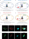
FIG. 1.
FIG. 1.
Role of IF1 in mitochondrial bio-energetics. ATP synthase activity under different conditions and mitochondrial localization of IF1 are depicted. Panels (a) and (b) illustrate the ATP synthase activity in normally respiting mitochondria, in the presence (a) of absence (b) of IF1. The electrons derived from biological oxidations are transferred to the respiratory chain complex to reduce oxygen and generate the electrochemical proton gradient across the inner membrane known as the PMF. ΔΨm serves the ATP synthase to produce ATP from ADP and inorganic phosphate. Mechanistically in fully active mitochondria, the ATP synthesis is driven by the influx of H+ into the mitochondrial matrix (a). During normoxic conditions, IF1 can partially inhibit the synthetic activity of the ATP synthase, resulting in an increase ΔΨm (b). Panels (c) and (d) illustrate the ATP synthase activity under hypoxic conditions or when the ETC system is dysfunctional and ΔΨm is not maintained. Under these conditions, the ATP synthase can reverse, consuming ATP and acting as a proton translocating ATPase to preserve ΔΨm (c). IF1 inhibits the ATP synthase hydrolytic activity, preserving cellular ATP at the expense of the ΔΨm (d). Panel (e) and (f) illustrate the co-localization of IF1 (green, rabbit polyclonal IF1 antibody (56) and Alexa Fluor 488-rabbit conjugated secondary antibody) with mitochondria (MitoTracker Red, Molecular Probes) in permeabilized HeLa cell line (e) and HCT-116 human colon cancer cell line (f). The nuclei were stained with DAPI (blue). Cell images were captured with an LSM 780 Zeiss confocal microscope. ΔΨm, mitochondrial membrane potential; ETC, electron transport chain; IF1, inhibitory factor 1; IMM, inner mitochondrial membrane; OMM, outer mitochondrial membrane; PMF, proton motive force; IMS, intermembrane space. Color images are available online.
FIG. 2.
FIG. 2.
IF1 isoforms. Scheme showing the domains of the IF1 isoforms; all three isoforms are produced as precursors, having an MTS that is cleaved on entering the mitochondria and releases the mature protein; Isoform 1 contains a MIS, necessary for the inhibition of the ATP synthase; Isoform 1 can be phosphorylated at Serine39 by mitochondrial protein kinase A; The Site H49K indicates the site-directed mutagenesis—when histidine49 is changed by lysine, IF1-H49K is obtained, which is active even at pH >6.7; IF1 isoform 1 can be acetylated at Lysine57, 83, and 90; Isoform 1 has an oligomerization domain—residues 57–69, a dimerization domain—residues 62–106 and a Calmodulin-Binding Motif residues 58–67; The precursor of Isoform 2 has an MTS, an incomplete MIS, and an isoform 2 specific domain; The precursor of Isoform 3 has an MTS and an incomplete MIS; Amino acid abbreviation: M-Methionine, A-Alanine, V-Valine, T-Threonine, L-Leucine, R-Arginine, W-Tryptophan, G-Glycine, Q-Glutamine, F-Phenylalanine, D-Aspartic acid, S-Serine, E-Glutamic acid, N-Asparagine, I-Isoleucine, K-Lysine, Y-Tyrosine, H-Histidine. MIS, minimal inhibitory sequence; MTS, mitochondrial targeting sequence. Color images are available online.
FIG. 3.
FIG. 3.
IF1-mediated ROS signaling in nuclear reprogramming and tumor development. Metabolic reprogramming from OXPHOS to aerobic glycolysis through IF1-mediated inhibition of ATP synthase; this process induces mild ROS that activate NF-κB pathway and Bcl-xl, preventing apoptosis. Bcl-xl, B cell lymphoma-extra large; NF-κB, nuclear factor-κB; OXPHOS, oxidative phosphorylation; ROS, reactive oxygen species. Color images are available online.
FIG. 4.
FIG. 4.
IF1 and mitochondrial fitness. (a) Cellular adaptation to stress and tumorigenesis—IF1-mediated mild mitochondrial ROS production has a protective role by promoting stress adaptation, survival, and proliferation through the activation of the NF-κB pathway, Nrf2 pathway, Bcl-xl, AMPK, and Akt; this mechanism can act as a physiological cellular response, but it can contribute to tumorigenesis. (b) Metabolic by-products: NAD+/NADH redox state and α-ketoglutarate: In primary human skeletal cells that overexpressed IF, the decreased ratio of NAD+/NADH was observed, together with a decrease in the cellular level of α-ketoglutarate and impaired fatty acid oxidation. (c) Mitochondrial cristae remodeling—IF1 can have a beneficial role in the density of mitochondrial cristae by promoting the dimerization of the ATP synthase; however, several studies disprove this hypothesis. (d) Mitophagy—Under conditions of mitochondrial depolarization induced by CCCP (a widely used inducer of mitophagy), the hydrolytic activity of ATP synthase promoted the loss of ΔΨm and subsequent PINK1 accumulation and Parkin recruitment, followed by mitophagy; this process is inhibited in the absence of IF1 in cells. Akt, protein kinase b; AMPK, 5′ AMP-activated protein kinase; CCCP, carbonyl cyanide m-chlorophenyl hydrazone; Nrf2, NF-E2 p45-related factor 2; PINK1, phosphatase and tensin homolog (PTEN)-induced kinase 1; PTEN, phosphatase and tensin homolog. Color images are available online.
FIG. 5.
FIG. 5.
Functions of cell surface ATP synthase (ecto-F1-ATPase). The ecto-F1-ATPase is present on the plasma membrane of different cell types, possibly through a trafficking secretory pathway after the assembly in the mitochondria. Similarly, IF1 might be possibly released into the extracellular environment. Under physiological conditions, ecto-F1-ATPase only has a hydrolytic function, converting ATP in ADP. IF1 inhibits this action, whereas apoA-I stimulates it. The generated ADP can activate the P2Y1 receptor present on the plasma membrane of endothelial cells and it promotes cell survival and NO production. The extracellular ADP can also activate the P2Y13 receptor present on the plasma membrane of hepatocytes and promote HDL endocytosis; the cholesterol from HDL will eventually be removed via biliary lipid secretion. Certain studies have attributed a role in tumorigenesis to the ecto-F1-ATPase based on the possible (?) production of extracellular ATP achieved with a local inverse H+ gradient generated by the activity of an ectopic respiratory chain. The ecto-F1-ATPase has been associated with tumor angiogenesis due to its presence on endothelial cells and its inhibition by angiostatin, a protein with anti-angiogenic properties. By inhibiting the ectopic enzyme, angiostatin can reduce the amount of ATP, lower the intracellular pH, and reduce cell survival and proliferation. In tumor cells, Vγ9/Vδ2 T lymphocytes recognize ecto-F1-ATPase on the surface through their TCR, and apoA-I enhances the cellular response. The ecto-F1-ATPase interacts with MHC Class I molecules on the surface of nontumor cells via NKR and protects them from Vγ9/Vδ2 T lymphocyte recognition. Upper left inset: Apolipoprotein apoA-I could contribute to ecto-IF1 release in extracellular medium of hepatocytes. HepG2 cells (3 × 106 per condition) were stabilized 1 h in serum-free medium and then incubated 20 min at 4°C with or without A-I (0, 10, and 50 μg/mL). The presence of IF1 was then analyzed in extracellular medium (eIF1) by immunoprecipitation with anti-IF1 antibody and Western blot (56). apoA-I, apolipoprotein A-I; MHC, major histocompatibility complex; NKR, natural killer receptors; TCR, T cell receptors. Color images are available online.
FIG. 6.
FIG. 6.
The potential involvement of IF1 in several diseases. Cancer: IF1 is overexpressed in various types of carcinomas, being a marker of poor (ovary, lung, colon, breast carcinomas) or favorable (colon, breast carcinomas) prognosis. The IF1 could induce metabolic reprogramming in cancer, by inhibiting OXPHOS and favoring aerobic glycolysis. Neurodegenerative diseases: IF1 could influence neurodegenerative disease progression by reducing extracellular ATP. The IF1 overexpression was associated with increased cognitive functions and protection from neurotoxicity during neuronal overstimulation. Cardiovascular diseases: IF1 has a positive impact in myocardial ischemia–reperfusion by preventing ATP depletion and cellular death. The IF1 promotes cardiac hypertrophy and can favor atherogenesis by inhibiting the atheroprotective activities of ecto-F1-ATPase present on hepatocytes (reverse cholesterol transport) and endothelial cells (NO production, cell survival). Metabolic diseases (diabetes, obesity): In the pancreas (INS-1E cells), IF1 decreases glucose-stimulated insulin secretion and glucose sensitivity, whereas in skeletal muscle, IF1 can stimulate TNF-α secretion and decrease β-oxidation. Dash arrows represent the inhibition of the ecto-F1-ATP synthase by IF1. Full arrows represent the inhibition of mitochondrial ATP synthase by IF1. TNFα, tumor necrosis factor alpha. Color images are available online.

References

    1. Allen DG, Morris PG, Orchard CH, and Pirolo JS. A nuclear magnetic resonance study of metabolism in the ferret heart during hypoxia and inhibition of glycolysis. J Physiol 361: 185–204, 1985. - PMC - PubMed
    1. Asmann YW, Stump CS, Short KR, Coenen-Schimke JM, Guo ZK, Bigelow ML, and Nair KS. Skeletal muscle mitochondrial functions, mitochondrial DNA copy numbers, and gene transcript profiles in type 2 diabetic and nondiabetic subjects at equal levels of low or high insulin and euglycemia. Diabetes 55: 3309–3319, 2006. - PubMed
    1. Bae TJ, Kim MS, Kim JW, Kim BW, Choo HJ, Lee JW, Kim KB, Lee CS, Kim JH, Chang SY, Kang CY, Lee SW, and Ko YG. Lipid raft proteome reveals ATP synthase complex in the cell surface. Proteomics 4: 3536–3548, 2004. - PubMed
    1. Bakula D and Scheibye-Knudsen M. MitophAging: mitophagy in aging and disease. Front Cell Dev Biol 8: 239, 2020. - PMC - PubMed
    1. Barazzuol L, Giamogante F, Brini M, and Calì T. PINK1/parkin mediated mitophagy, Ca2+ signalling, and er-mitochondria contacts in Parkinson's disease. Int J Mol Sci 21: 1772, 2020. - PMC - PubMed

Publication types

MeSH terms

Substances

LinkOut - more resources