. 2022 Jun 3;145(5):1773-1784.
doi: 10.1093/brain/awab372.

Cholinergic white matter pathways in dementia with Lewy bodies and Alzheimer's disease

Affiliations

Cholinergic white matter pathways in dementia with Lewy bodies and Alzheimer's disease

Julia Schumacher et al. Brain. .

Abstract

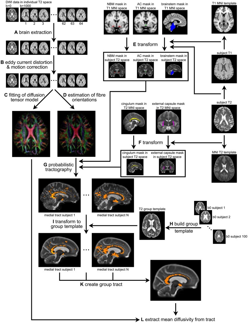
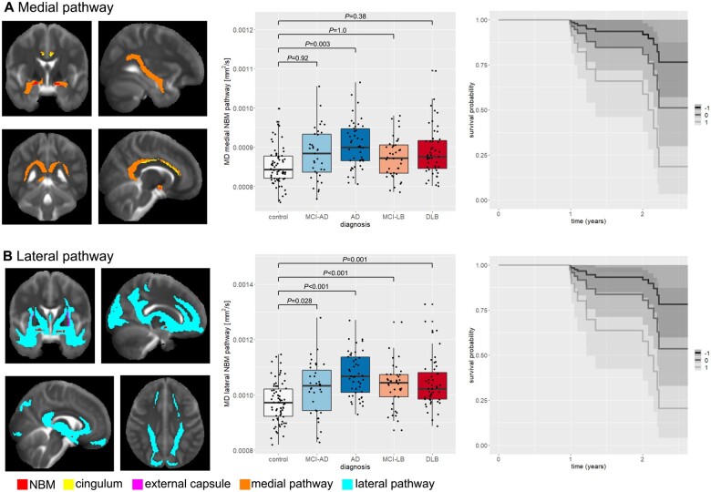
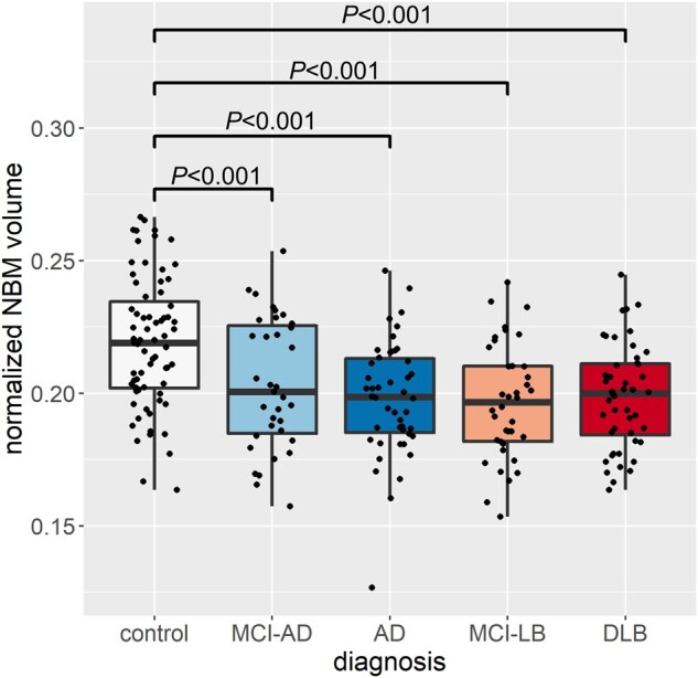
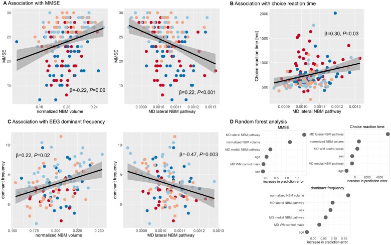
Patients who have dementia with Lewy bodies and Alzheimer's disease show early degeneration of the cholinergic nucleus basalis of Meynert. However, how white matter projections between the nucleus basalis of Meynert and the cortex are altered in neurodegenerative disease is unknown. Tractography of white matter pathways originating from the nucleus basalis of Meynert was performed using diffusion-weighted imaging in 46 patients with Alzheimer's disease dementia, 48 with dementia with Lewy bodies, 35 with mild cognitive impairment with Alzheimer's disease, 38 with mild cognitive impairment with Lewy bodies and 71 control participants. Mean diffusivity of the resulting pathways was compared between groups and related to cognition, attention, functional EEG changes and dementia conversion in the mild cognitive impairment groups. We successfully tracked a medial and a lateral pathway from the nucleus basalis of Meynert. Mean diffusivity of the lateral pathway was higher in both dementia and mild cognitive impairment groups than controls (all P < 0.03). In the patient groups, increased mean diffusivity of this pathway was related to more impaired global cognition (β = -0.22, P = 0.06) and worse performance on an attention task (β = 0.30, P = 0.03). In patients with mild cognitive impairment, loss of integrity of both nucleus basalis of Meynert pathways was associated with increased risk of dementia progression [hazard ratio (95% confidence interval), medial pathway: 2.51 (1.24-5.09); lateral pathway: 2.54 (1.24-5.19)]. Nucleus basalis of Meynert volume was reduced in all clinical groups compared to controls (all P < 0.001), but contributed less strongly to cognitive impairment and was not associated with attention or dementia conversion. EEG slowing in the patient groups as assessed by a decrease in dominant frequency was associated with smaller nucleus basalis of Meynert volumes (β = 0.22, P = 0.02) and increased mean diffusivity of the lateral pathway (β = -0.47, P = 0.003). We show that degeneration of the cholinergic nucleus basalis of Meynert in Alzheimer's disease and dementia with Lewy bodies is accompanied by an early reduction in integrity of white matter projections that originate from this structure. This is more strongly associated with cognition and attention than the volume of the nucleus basalis of Meynert itself and might be an early indicator of increased risk of dementia conversion in people with mild cognitive impairment.

Keywords: basal forebrain; diffusion-weighted imaging; mild cognitive impairment; nucleus basalis of Meynert; probabilistic tractography.


Figures

Figure 1
Figure 1
Overview of NBM white matter tract estimation. (A) Brain extraction of diffusion-weighted data using FSL’s bet function. (B) Correction for eddy currents and head motion using FSL’s eddy tool. (C) Fitting of the diffusion tensor model using FSL’s dtifit. (D) Estimation of fibre orientations in each voxel using FSL’s BedpostX with a ball-and-stick model with three fibres per voxel. (E) Transformation of region of interest from T1 MNI space to individual subject T2 space. (F) Transformation of region of interest from standard T2 space to individual subject T2 space. (G) Probabilistic tractography with FSL’s ProbtrackX using the NBM region of interest as seed, the cingulum and external capsule region of interest as waypoint masks, and the brainstem and anterior commissure as exclusion masks. (H) Building of unbiased group template from 100 randomly selected b0 images. (I) Transformation of tracts from individual subject T2 space to the unbiased group template. (K) Estimation of group tracts for the medial and lateral NBM pathways. (L) Extraction of diffusion parameters from the estimated tracts. AC = anterior commissure.
Figure 2
Figure 2
Group comparison of NBM pathway integrity. (A) Medial NBM pathway (shown in orange) estimated with the NBM region of interest as seed (shown in red), the cingulum as waypoint mask (shown in yellow), and brainstem and anterior commissure as exclusion masks (not shown). (B) Lateral NBM pathway (shown in cyan) estimated with the NBM region of interest as seed (shown in red), the external capsule as waypoint mask (shown in pink), and the brainstem and anterior commissure as exclusion masks (not shown). In each box plot the central line corresponds to the sample median, the upper and lower border of the box represent the 25th and 75th percentile, respectively, and the length of the whiskers corresponds to 1.5 times the interquartile range. P-values result from pairwise post hoc tests with Bonferroni correction for multiple comparisons. All P-values for comparisons that are not shown are >0.05. The plots on the right show survival probabilities in MCI without dementia given a standardized (A) medial and (B) lateral pathway mean diffusivity of −1, 0, or +1 (representative z-scores with 0 being the mean, +1 being 1 standard deviation (SD) higher and −1 being 1 SD lower than mean diffusivity). AD = Alzheimer’s disease dementia; MD = mean diffusivity.
Figure 3
Figure 3
Group comparison of normalized NBM volume. In each box plot the central line corresponds to the sample median, the upper and lower border of the box represent the 25th and 75th percentile, respectively, and the length of the whiskers corresponds to 1.5 times the interquartile range. P-values result from pairwise post hoc tests with Bonferroni correction for multiple comparisons. All P-values for comparisons that are not shown are >0.05. AD = Alzheimer’s disease dementia.
Figure 4
Figure 4
Association with cognitive and attentional performance. Association between mean diffusivity (MD) along NBM pathways and (A) MMSE as a measure of overall cognition, (B) choice reaction time and (C) EEG dominant frequency. β-values result from a multiple linear regression analysis including mean diffusivity along the two NBM pathways, normalized NBM volume, diagnosis, age, sex and mean diffusivity from the white matter control mask as predictors. Grey shaded areas correspond to 95% confidence intervals. (D) Results from the random forest analysis showing the conditional feature importance (i.e. increase in prediction error when permuting a feature on a grid defined by the other features in the model). Higher values indicate higher importance of a feature in predicting the response variable (choice reaction time, MMSE and EEG dominant frequency). AD = Alzheimer’s disease dementia; MD = mean diffusivity; WM = white matter.
Figure 5
Figure 5
Correlation structure between different predictor variables. Scatterplots (bottom left) and Pearson’s correlation values (top right) between mean diffusivity from the two NBM pathways, NBM volume and diffusivity of the white matter control mask, across all diagnostic groups and for each group separately. Significant correlations (P < 0.05) are marked with an asterisk. AD = Alzheimer’s disease dementia; MD = mean diffusivity; WM = white matter.

References

    1. Bohnen NI, Grothe MJ, Ray NJ, Müller MLTM, Teipel SJ.. Recent advances in cholinergic imaging and cognitive decline—revisiting the cholinergic hypothesis of dementia. Curr Geriatr Reports. 2018;7(1):1–11. - PMC - PubMed
    1. Gratwicke J, Kahan J, Zrinzo L, et al. . The nucleus basalis of Meynert: A new target for deep brain stimulation in dementia? Neurosci Biobehav Rev. 2013;37(10 Pt 2):2676–2688. - PubMed
    1. Gratwicke J, Jahanshahi M, Foltynie T.. Parkinson’s disease dementia: A neural networks perspective. Brain. 2015;138(Pt 6):1454–1476. - PMC - PubMed
    1. Mesulam M-M. Cholinergic circuitry of the human nucleus basalis and its fate in Alzheimer’s disease. J Comp Neurol. 2013;521(18):4124–4144. - PMC - PubMed
    1. Colloby SJ, Elder GJ, Rabee R, O’Brien JT, Taylor J-P.. Structural grey matter changes in the substantia innominata in Alzheimer’s disease and dementia with Lewy bodies: A DARTEL-VBM study. Int J Geriatr Psychiatry. 2017;32(6):615–623. - PMC - PubMed

Publication types

MeSH terms

Substances