. 2021 Nov 19;203(24):e0045821.
doi: 10.1128/JB.00458-21. Epub 2021 Oct 4.

Heme-Dependent Siderophore Utilization Promotes Iron-Restricted Growth of the Staphylococcus aureus hemB Small-Colony Variant

Affiliations

Heme-Dependent Siderophore Utilization Promotes Iron-Restricted Growth of the Staphylococcus aureus hemB Small-Colony Variant

Izabela Z Batko et al. J Bacteriol. .

Abstract

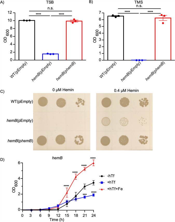
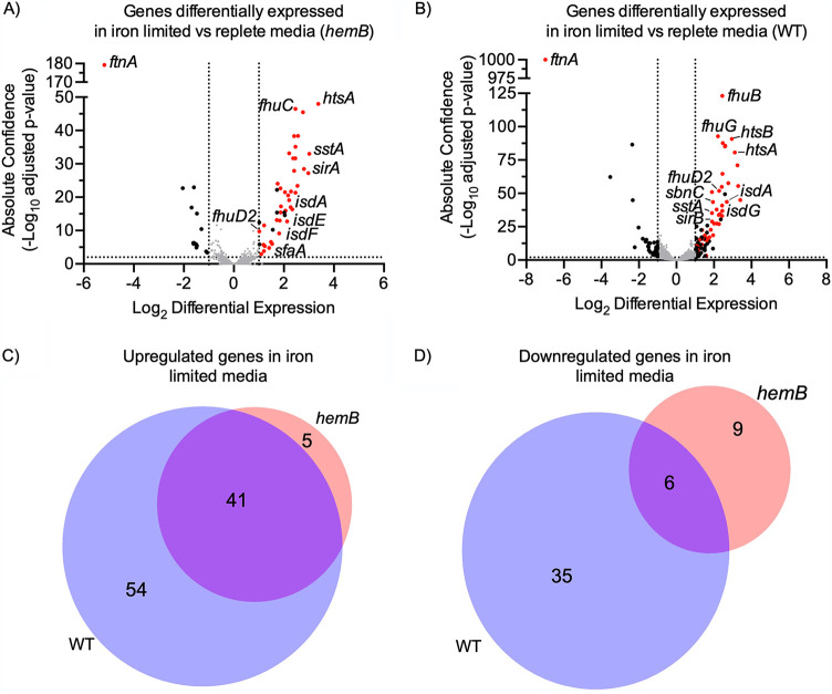
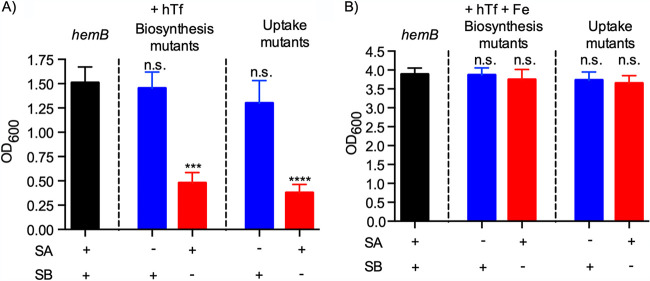
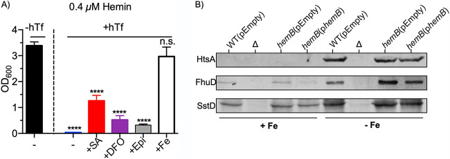
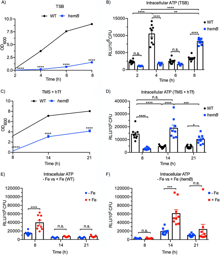
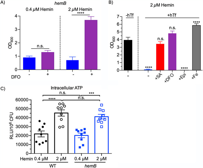
Respiration-deficient Staphylococcus aureus small-colony variants (SCVs) frequently cause persistent infections, which necessitates they acquire iron, yet how SCVs obtain iron remains unknown. To address this, we created a stable hemB mutant from S. aureus USA300 strain LAC. The hemB SCV utilized exogenously supplied hemin but was attenuated for growth under conditions of iron starvation. Transcriptome sequencing (RNA-seq) showed that both wild-type (WT) S. aureus and the hemB mutant sense and respond to iron starvation; however, growth assays show that the hemB mutant is defective for siderophore-mediated iron acquisition. Indeed, the hemB SCV demonstrated limited utilization of endogenous staphyloferrin B or exogenously provided staphyloferrin A, deferoxamine mesylate (Desferal), and epinephrine. Direct measurement of intracellular ATP in hemB and WT S. aureus revealed that both strains can generate comparable levels of ATP during exponential growth, suggesting defects in ATP production cannot account for the inability to efficiently utilize siderophores. Defective siderophore utilization by hemB bacteria was also evident in vivo, as administration of Desferal failed to promote hemB bacterial growth in every organ analyzed except for the kidneys. In support of the hypothesis that S. aureus accesses heme in kidney abscesses, in vitro analyses revealed that increased hemin availability enables hemB bacteria to utilize siderophores for growth when iron availability is restricted. Taken together, our data support the conclusion that hemin is used not only as an iron source itself but also as a nutrient that promotes utilization of siderophore-iron complexes. IMPORTANCE S. aureus small-colony variants (SCVs) are associated with chronic recurrent infection and worsened clinical outcome. SCVs persist within the host despite administration of antibiotics. This study yields insight into how S. aureus SCVs acquire iron, which during infection of a host is a difficult-to-acquire metal nutrient. Under hemin-limited conditions, hemB S. aureus is impaired for siderophore-dependent growth, and in agreement, murine infection indicates that hemin-deficient SCVs meet their nutritional requirement for iron through utilization of hemin. Importantly, we demonstrate that hemB SCVs rely upon hemin as a nutrient to promote siderophore utilization. Therefore, perturbation of heme biosynthesis and/or utilization represents a viable to strategy to mitigate the ability of SCV bacteria to acquire siderophore-bound iron during infection.

Keywords: SCV; heme; iron starvation; pathogenesis; siderophores.


Figures

FIG 1
FIG 1
Growth of S. aureus hemB SCV in rich and minimal media. Growth of S. aureus USA300 WT(pEmpty), hemB(pEmpty), and hemB(phemB) strains cultured in tryptic soy broth (TSB) (A) or Tris minimal succinate (TMS) (B) for 24 h. Data are plotted as the mean ± standard error of the mean (SEM) from three biological replicates. ****, P ≤ 0.0001 by one-way analysis of variance (ANOVA) with Tukey’s multiple comparisons; n.s., not significant. (C) Growth of S. aureus USA300 WT(pEmpty), hemB(pEmpty), and hemB(phemB) strains on TMS agar supplemented with either 0 μM (left panel) or 0.4 μM (right panel) hemin after 48 h of incubation at 37°C. (D) Growth over 24 h at 37°C of the hemB mutant cultured in TMS supplemented with 0.4 μM hemin and either no human transferrin (−hTf), 1.5 μM human transferrin (+hTf), or 1.5 μM human transferrin and 10 μM ferrous ammonium sulfate (+hTf+Fe). Data are plotted as the mean ± SEM from five biological replicates. ***, P ≤ 0.001, and ****, P ≤ 0.0001, by two-way ANOVA with Dunnett’s multiple comparisons.
FIG 2
FIG 2
Analysis of RNA expression in response to iron starvation by S. aureus WT and hemB strains. Shown are volcano plots of genes that were differentially expressed in iron-limited versus iron-replete media by either the S. aureus hemB SCV (A) or WT S. aureus (B). Significantly up- and downregulated genes (absolute confidence of >2 and |log2 differential expression| of >1) that are Fur regulated are colored red, and the remaining significantly regulated genes are colored black. (C and D) Comparison of the number of significantly upregulated (C) or significantly downregulated (D) genes in iron-limited media by WT and hemB S. aureus. Raw reads for RNA-seq data and lists of genes significantly up- and downregulated under all conditions can be found in Table S1.
FIG 3
FIG 3
Staphyloferrin B-mediated iron acquisition is required for optimal iron-starved growth of hemB SCV. Shown is growth of hemB S. aureus (black) or hemB S. aureus combined with mutation of SA (blue) or SB (red) iron acquisition systems. SA and SB biosynthetic and uptake mutations are indicated. Bacteria were cultured for 24 h in TMS supplemented with 0.4 μM hemin and 1.5 μM human transferrin (+hTf) (A) or 1.5 μM human transferrin and 10 μM ferrous ammonium sulfate (+hTf+Fe) (B). Data are plotted as the mean ± SEM from nine biological replicates from three independent experiments. ***, P ≤ 0.001, and ****, P ≤ 0.0001, by one-way ANOVA with Tukey’s multiple comparisons; n.s., not significant.
FIG 4
FIG 4
Siderophore utilization by S. aureus hemB is defective. Shown is growth of the staphyloferrin-deficient hemB sfa sbn mutant cultured for 24 h in TMS supplemented with 0.4 μM hemin. Medium contains either no human transferrin (−hTf) or 5 μM human transferrin (+hTf). Either 100 μM siderophore (staphyloferrin A [+SA], deferoxamine mesylate [+DFO], or epinephrine [+Epi]) or 30 μM ferrous ammonium sulfate (+Fe) was added to the iron-restricted medium. Data are plotted as the mean ± SEM from nine biological replicates from three independent experiments. ****, P ≤ 0.0001 by one-way ANOVA with Tukey’s multiple comparisons; n.s., not significant. (B) Western blots for detection of HtsA (31 kDa), FhuD2 (34 kDa), or SstD (38 kDa) expression by the WT(pEmpty), hemB(pEmpty), or hemB(phemB) strain. A negative control (Δ)—either an S. aureus htsA, fhuD2, or sstD mutant—was used in each respective Western blot. Whole-cell lysates were prepared from bacteria cultured for 24 h in TMS supplemented with 0.4 μM hemin and either 30 μM ferrous ammonium sulfate (+Fe) or 1.5 μM human transferrin (−Fe).
FIG 5
FIG 5
Intracellular ATP of an S. aureus hemB mutant. (A and B) Growth (A) and intracellular ATP levels (B) of WT and hemB S. aureus grown in TSB. (C and D) Growth (C) and intracellular ATP levels (D) of WT and hemB S. aureus grown in TMS supplemented with 0.4 μM hemin and 1.5 μM human transferrin (+hTf). (E and F) Intracellular ATP levels of WT S. aureus (E) or the hemB mutant (F) grown in TMS supplemented with 0.4 μM hemin, 1.5 μM hTf, and either 0 μM ferrous ammonium sulfate (−Fe) or 10 μM ferrous ammonium sulfate (+Fe). Data are plotted as the mean ± SEM from nine biological replicates from three independent experiments. *, P < 0.05, **, P < 0.01, ***, P < 0.001, and ****, P < 0.0001, by two-way ANOVA with Dunnett’s multiple comparisons for panels A and C or one-way ANOVA with Tukey’s posttest for panels B and D to F; n.s., not significant.
FIG 6
FIG 6
Niche-specific utilization of DFO by S. aureus hemB in vivo in a murine model of systemic infection. Mice were inoculated with the S. aureus USA300 WT or hemB mutant and received either DFO or vehicle control (see Materials and Methods). Forty-eight hours postinfection, mice were sacrificed, and the percentage of weight loss (A) and bacterial burden of the heart (B), kidneys (C), or liver (D) were determined. The limit of accurate detection is represented as a dashed line. Data are plotted as the mean ± SEM from at least 15 animals per group. *, P ≤ 0.05, **, P ≤ 0.01, ***, P ≤ 0.001, and ****, P ≤ 0.0001, by one-way ANOVA with Tukey’s posttest; n.s., not significant.
FIG 7
FIG 7
Siderophore utilization by S. aureus hemB is enhanced by additional hemin. (A) Growth of the hemB mutant cultured at 37°C for 24 h in iron-restricted TMS supplemented with either 0.4 or 2 μM hemin, as indicated. The medium contains 5 μM hTf to restrict iron and either 0 or 100 μM DFO to assess utilization of the hydroxamate-type siderophore. Data are plotted as the mean ± SEM from at least five biological replicates. ****, P ≤ 0.0001 by one-way ANOVA with Tukey’s multiple comparisons; n.s., not significant. (B) Growth of the staphyloferrin-deficient hemB sfa sbn mutant cultured at 37°C for 24 h in TMS supplemented with 2 μM hemin. The medium contains either no human transferrin (−hTf) or 5 μM human transferrin (+hTf). Either 100 μM siderophore (staphyloferrin A [+SA], deferoxamine mesylate [+DFO], or epinephrine [+Epi]) or 30 μM ferrous ammonium sulfate (+Fe) was added to the iron-restricted medium. (C) Intracellular ATP levels of WT and hemB S. aureus grown to exponential phase in TMS supplemented with either 0.4 or 2 μM hemin. Data are plotted as the mean ± SEM from nine biological replicates from three independent experiments. ***, P ≤ 0.001, and ****, P ≤ 0.0001, by one-way ANOVA with Tukey’s multiple comparisons.

Similar articles

Cited by

References

    1. Turner NA, Sharma-Kuinkel BK, Maskarinec SA, Eichenberger EM, Shah PP, Carugati M, Holland TL, Fowler VG, Jr.. 2019. Methicillin-resistant Staphylococcus aureus: an overview of basic and clinical research. Nat Rev Microbiol 17:203–218. 10.1038/s41579-018-0147-4. - DOI - PMC - PubMed
    1. Tong SYC, Davis JS, Eichenberger E, Holland TL, Fowler VG. 2015. Staphylococcus aureus infections: epidemiology, pathophysiology, clinical manifestations, and management. Clin Microbiol Rev 28:603–661. 10.1128/CMR.00134-14. - DOI - PMC - PubMed
    1. Boucher H, Miller LG, Razonable RR. 2010. Serious infections caused by methicillin-resistant Staphylococcus aureus. Clin Infect Dis 51(Suppl 2):S183–S197. 10.1086/653519. - DOI - PubMed
    1. Kochanek KD, Xu J, Murphy SL, Miniño AM, Kung H-C. 2012. Deaths: final data for 2009. Natl Vital Stat Rep 60:1–116. - PubMed
    1. van Hal SJ, Jensen SO, Vaska VL, Espedido BA, Paterson DL, Gosbell IB. 2012. Predictors of mortality in Staphylococcus aureus bacteremia. Clin Microbiol Rev 25:362–386. 10.1128/CMR.05022-11. - DOI - PMC - PubMed

Publication types

MeSH terms

Grants and funding