

Y chromosome functions in mammalian spermatogenesis
- PMID: 34606444
- PMCID: PMC8489898
- DOI: 10.7554/eLife.67345
Y chromosome functions in mammalian spermatogenesis
Abstract
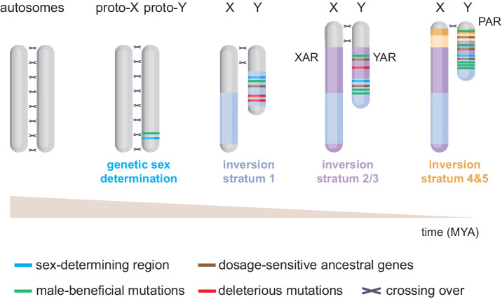
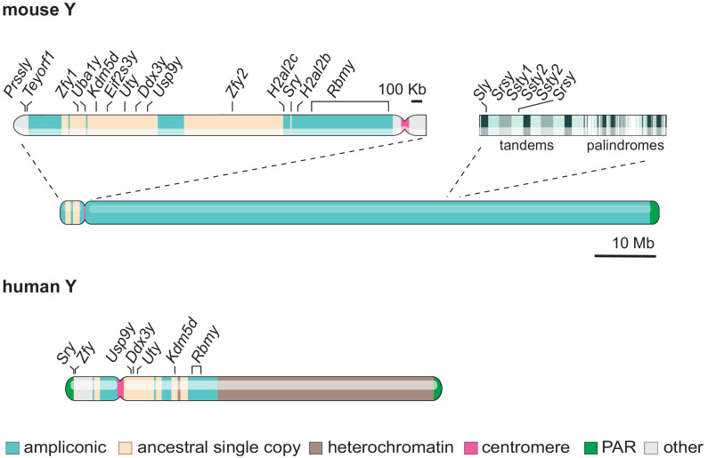
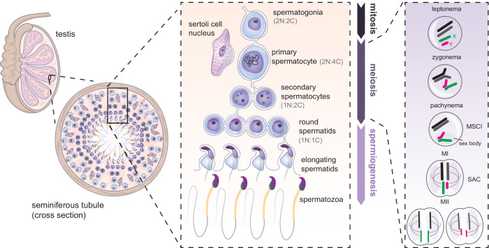
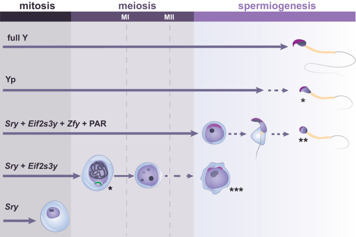
The mammalian Y chromosome is critical for male sex determination and spermatogenesis. However, linking each Y gene to specific aspects of male reproduction has been challenging. As the Y chromosome is notoriously hard to sequence and target, functional studies have mostly relied on transgene-rescue approaches using mouse models with large multi-gene deletions. These experimental limitations have oriented the field toward the search for a minimum set of Y genes necessary for male reproduction. Here, considering Y-chromosome evolutionary history and decades of discoveries, we review the current state of research on its function in spermatogenesis and reassess the view that many Y genes are disposable for male reproduction.
Keywords: Y chromosome; developmental biology; fertility; spermatogenesis.
© 2021, Subrini and Turner.
Conflict of interest statement
JS, JT No competing interests declared
Figures
Similar articles
-
The Biology and Evolution of Mammalian Y Chromosomes.Annu Rev Genet. 2015;49:507-27. doi: 10.1146/annurev-genet-112414-055311. Epub 2015 Oct 6. Annu Rev Genet. 2015. PMID: 26442847 Review.
-
Mammalian Sex Chromosome Structure, Gene Content, and Function in Male Fertility.Annu Rev Anim Biosci. 2019 Feb 15;7:103-124. doi: 10.1146/annurev-animal-020518-115332. Epub 2018 Nov 9. Annu Rev Anim Biosci. 2019. PMID: 30412673 Review.
-
The role of Y-encoded genes in mammalian spermatogenesis.Semin Cell Dev Biol. 1998 Aug;9(4):423-32. doi: 10.1006/scdb.1998.0228. Semin Cell Dev Biol. 1998. PMID: 9813189 Review.
-
Mammalian Y chromosome evolution and the male-specific functions of Y chromosome-borne genes.Rev Reprod. 1999 May;4(2):101-9. doi: 10.1530/ror.0.0040101. Rev Reprod. 1999. PMID: 10357097 Review.
-
The Y chromosome.Reprod Biomed Online. 2002 Jul-Aug;5(1):22-5. doi: 10.1016/s1472-6483(10)61592-1. Reprod Biomed Online. 2002. PMID: 12470541
Cited by
-
Conserved genes regulating human sex differentiation, gametogenesis and fertilization.J Transl Med. 2024 May 19;22(1):473. doi: 10.1186/s12967-024-05162-2. J Transl Med. 2024. PMID: 38764035 Free PMC article. Review.
-
Where Are the Formerly Y-linked Genes in the Ryukyu Spiny Rat that has Lost its Y Chromosome?Genome Biol Evol. 2024 Mar 2;16(3):evae046. doi: 10.1093/gbe/evae046. Genome Biol Evol. 2024. PMID: 38478711 Free PMC article.
-
PRAMEY: A Bovid-Specific Y-Chromosome Multicopy Gene Is Highly Related to Postnatal Testicular Growth in Hu Sheep.Animals (Basel). 2022 Sep 12;12(18):2380. doi: 10.3390/ani12182380. Animals (Basel). 2022. PMID: 36139240 Free PMC article.
-
Y chromosome introgression between deeply divergent primate species.Nat Commun. 2024 Nov 29;15(1):10398. doi: 10.1038/s41467-024-54719-8. Nat Commun. 2024. PMID: 39613758 Free PMC article.
-
A researcher's guide to studying sex differences in immune aging.Trends Mol Med. 2025 Aug;31(8):702-717. doi: 10.1016/j.molmed.2025.01.005. Epub 2025 Jan 29. Trends Mol Med. 2025. PMID: 39884873 Review.
References
Publication types
MeSH terms
Grants and funding
LinkOut - more resources
Full Text Sources
Research Materials
