Autoantibodies Present in Hidradenitis Suppurativa Correlate with Disease Severity and Promote the Release of Proinflammatory Cytokines in Macrophages
- PMID: 34606886
- PMCID: PMC8860851
- DOI: 10.1016/j.jid.2021.07.187
Autoantibodies Present in Hidradenitis Suppurativa Correlate with Disease Severity and Promote the Release of Proinflammatory Cytokines in Macrophages
Abstract
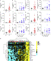
Hidradenitis suppurativa (HS), also known as acne inversa, is a debilitating inflammatory skin disorder that is characterized by nodules that lead to the development of connected tunnels and scars as it progresses from Hurley stages I to III. HS has been associated with several autoimmune diseases, including inflammatory bowel disease and spondyloarthritis. We previously reported dysregulation of humoral immune responses in HS, characterized by elevated serum total IgG, B-cell activation, and antibodies recognizing citrullinated proteins. In this study, we characterized IgG autoreactivity in HS sera and lesional skin compared with those in normal healthy controls using an array-based high-throughput autoantibody screening. The Cy3-labeled anti-human assay showed the presence of autoantibodies against nuclear antigens, cytokines, cytoplasmic proteins, extracellular matrix proteins, neutrophil proteins, and citrullinated antigens. Most of these autoantibodies were significantly elevated in stages II‒III in HS sera and stage III in HS skin lesions compared with those of healthy controls. Furthermore, immune complexes containing both native and citrullinated versions of antigens can activate M1 and M2 macrophages to release proinflammatory cytokines such as TNF-α, IL-8, IL-6, and IL-12. Taken together, the identification of specific IgG autoantibodies that recognize circulating and tissue antigens in HS suggests an autoimmune mechanism and uncovers putative therapeutic targets.
Copyright © 2021 The Authors. Published by Elsevier Inc. All rights reserved.
Figures


Similar articles
-
Neutrophil extracellular traps, B cells, and type I interferons contribute to immune dysregulation in hidradenitis suppurativa.Sci Transl Med. 2019 Sep 4;11(508):eaav5908. doi: 10.1126/scitranslmed.aav5908. Sci Transl Med. 2019. PMID: 31484788 Free PMC article.
-
Two Cases of Hidradenitis Suppurativa Treated with Adalimumab at the Department of Dermatology and Venereology, Clinical Hospital Mostar.Acta Dermatovenerol Croat. 2021 Jul;29(2):108-110. Acta Dermatovenerol Croat. 2021. PMID: 34477078
-
Neutrophil extracellular traps activate Notch-γ-secretase signaling in hidradenitis suppurativa.J Allergy Clin Immunol. 2025 Jan;155(1):188-198. doi: 10.1016/j.jaci.2024.09.001. Epub 2024 Sep 17. J Allergy Clin Immunol. 2025. PMID: 39265876
-
Hidradenitis suppurativa.Med Clin (Barc). 2024 Feb 23;162(4):182-189. doi: 10.1016/j.medcli.2023.09.018. Epub 2023 Nov 13. Med Clin (Barc). 2024. PMID: 37968174 Review. English, Spanish.
-
Evidence-based approach to the treatment of hidradenitis suppurativa/acne inversa, based on the European guidelines for hidradenitis suppurativa.Rev Endocr Metab Disord. 2016 Sep;17(3):343-351. doi: 10.1007/s11154-016-9328-5. Rev Endocr Metab Disord. 2016. PMID: 26831295 Free PMC article. Review.
Cited by
-
Autoimmune, Autoinflammatory Disease and Cutaneous Malignancy Associations with Hidradenitis Suppurativa: A Cross-Sectional Study.Am J Clin Dermatol. 2024 May;25(3):473-484. doi: 10.1007/s40257-024-00844-5. Epub 2024 Feb 9. Am J Clin Dermatol. 2024. PMID: 38337127
-
CD2 expressing innate lymphoid and T cells are critical effectors of immunopathogenesis in hidradenitis suppurativa.Proc Natl Acad Sci U S A. 2024 Nov 26;121(48):e2409274121. doi: 10.1073/pnas.2409274121. Epub 2024 Nov 19. Proc Natl Acad Sci U S A. 2024. PMID: 39560648 Free PMC article.
-
Hidradenitis suppurativa: key insights into treatment success and failure.J Clin Invest. 2024 Nov 1;134(21):e186744. doi: 10.1172/JCI186744. J Clin Invest. 2024. PMID: 39484718 Free PMC article. No abstract available.
-
B cells in non-lymphoid tissues.Nat Rev Immunol. 2025 Jul;25(7):483-496. doi: 10.1038/s41577-025-01137-6. Epub 2025 Feb 5. Nat Rev Immunol. 2025. PMID: 39910240 Review.
-
Skin immune-mesenchymal interplay within tertiary lymphoid structures promotes autoimmune pathogenesis in hidradenitis suppurativa.Immunity. 2024 Dec 10;57(12):2827-2842.e5. doi: 10.1016/j.immuni.2024.11.010. Immunity. 2024. PMID: 39662091
References
-
- Abbate A, Van Tassell BW, Biondi-Zoccai GG. Blocking interleukin-1 as a novel therapeutic strategy for secondary prevention of cardiovascular events. BioDrugs 2012;26:217–33. - PubMed
-
- Assan F, Gottlieb J, Tubach F, Lebbah S, Guigue N, Hickman G, et al. Anti-Saccharomyces cerevisiae IgG and IgA antibodies are associated with systemic inflammation and advanced disease in hidradenitis suppurativa. J Allergy Clin Immunol 2020;146. 452–5.e5. - PubMed
-
- Bodaño A, González A, Ferreiros-Vidal I, Balada E, Ordi J, Carreira P, et al. Association of a non-synonymous single-nucleotide polymorphism of DNASEI with SLE susceptibility. Rheumatology (Oxford) 2006;45:819–23. - PubMed
-
- Bonaventura A, Montecucco F. Inflammation and pericarditis: are neutrophils actors behind the scenes? J Cell Physiol 2019;234:5390–8. - PubMed
Publication types
MeSH terms
Substances
Grants and funding
LinkOut - more resources
Full Text Sources
Medical

