

Duodenal Mucosal Barrier in Functional Dyspepsia
- PMID: 34607017
- PMCID: PMC8975906
- DOI: 10.1016/j.cgh.2021.09.029
Duodenal Mucosal Barrier in Functional Dyspepsia
Abstract
Background & aims: In addition to gastric sensorimotor dysfunctions, functional dyspepsia (FD) is also variably associated with duodenal micro-inflammation and epithelial barrier dysfunction, the pathogenesis and clinical significance of which are unknown. Our hypothesis was that miRNAs and/or inflammation degrade epithelial barrier proteins, resulting in increased duodenal mucosal permeability in FD.
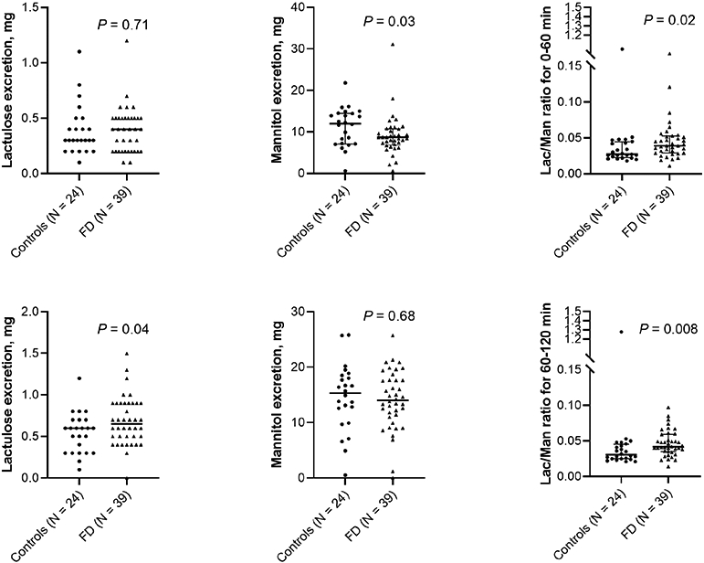
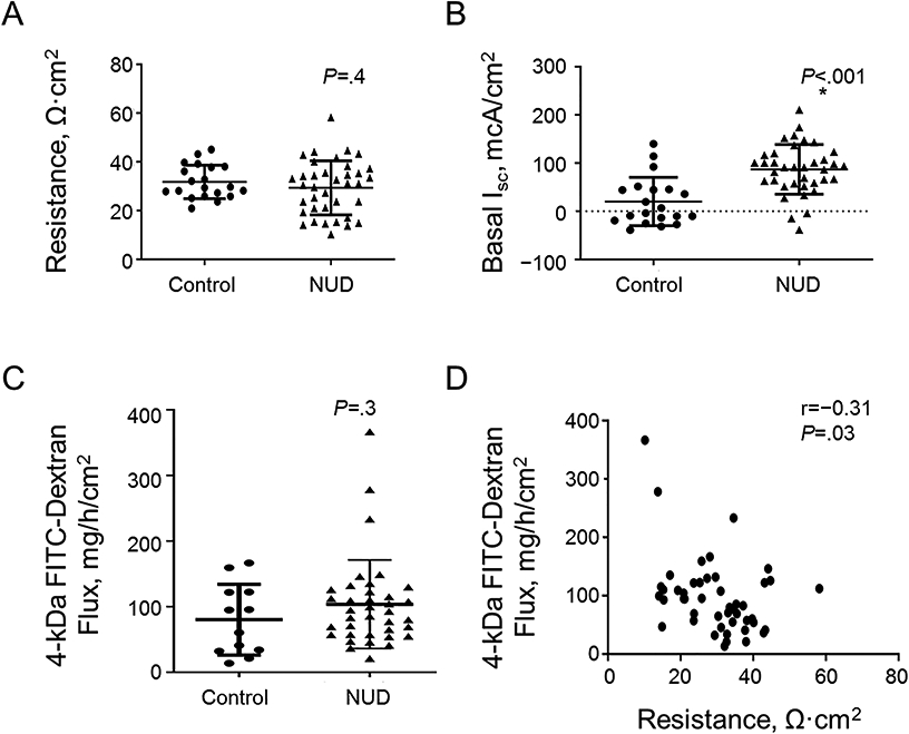
Methods: We compared the duodenal mucosal gene expression and miRNAs, in vivo permeability (lactulose-mannitol excretion between 0 and 60 and 60 and 120 minutes after saccharide ingestion), ex vivo assessments (transmucosal resistance, fluorescein isothiocyanate [FITC]-dextran flux, and basal ion transport), and duodenal histology (light and electron microscopy) in 40 patients with FD and 24 controls.
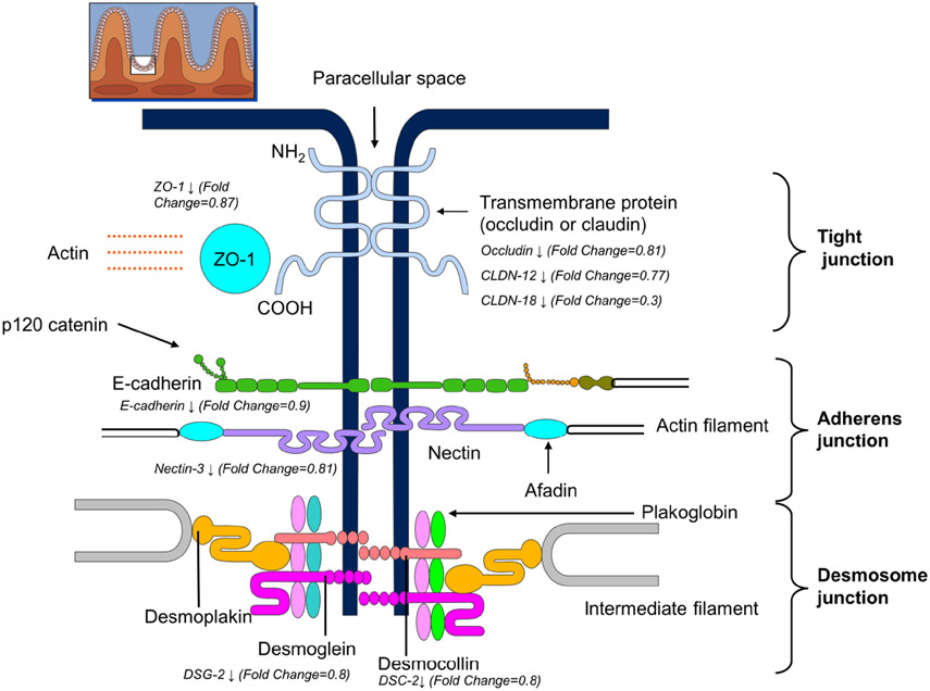
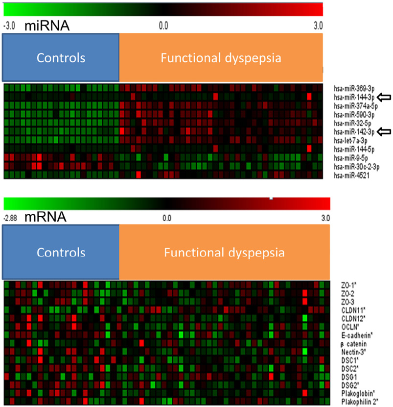
Results: Compared with controls, the mRNA expression of several barrier proteins (zonula occludens-1, occludin, claudin-12, and E-cadherin) was modestly reduced (ie, a fold change of 0.8-0.85) in FD with increased expression of several miRNAs (eg, miR-142-3p and miR-144-3-p), which suppress these genes. The urinary lactulose excretion and the lactulose:mannitol ratio between 60 and 120 minutes were greater in FD than in controls (P < .05). The FITC-dextran flux, which reflects paracellular permeability, was inversely correlated (r = -0.32, P = .03) with transmucosal resistance and directly correlated (r = 0.4, P = .02) with lactulose:mannitol ratio. Other parameters (mucosal eosinophils, intraepithelial lymphocytes, and mast cells, transmucosal resistance, FITC-dextran flux, average intercellular distance, and proportion of dilated junctions) were not significantly different between groups.
Conclusions: In FD, there is a modest reduction in the expression of several duodenal epithelial barrier proteins, which may be secondary to up-regulation of regulatory miRNAs, and increased small intestinal permeability measured in vivo.
Keywords: Duodenal Epithelial Permeability; Inflammation; Intestinal Barrier; Tight Junction Gene Expression.
Copyright © 2022 AGA Institute. Published by Elsevier Inc. All rights reserved.
Conflict of interest statement
Figures
Comment in
-
Duodenal Barrier and Inflammation in Dyspepsia: God is in the Details.Clin Gastroenterol Hepatol. 2022 Oct;20(10):2411-2413. doi: 10.1016/j.cgh.2021.11.005. Epub 2021 Nov 13. Clin Gastroenterol Hepatol. 2022. PMID: 34785357 No abstract available.
References
-
- Vanheel H, Carbone F, Valvekens L, et al. Pathophysiological Abnormalities in Functional Dyspepsia Subgroups According to the Rome III Criteria. American Journal of Gastroenterology 2017;112:132–140. - PubMed
-
- Talley NJ, Walker MM, Aro P, et al. Non-ulcer dyspepsia and duodenal eosinophilia: an adult endoscopic population-based case-control study. Clin Gastroenterol Hepatol 2007;5:1175–83. - PubMed
-
- Vanheel H, Vicario M, Vanuytsel T, et al. Impaired duodenal mucosal integrity and low-grade inflammation in functional dyspepsia. Gut 2014;63:262–71. - PubMed
-
- Du L, Chen B, Kim JJ, et al. Micro-inflammation in functional dyspepsia: A systematic review and meta-analysis. Neurogastroenterology & Motility 2018;30:e13304. - PubMed
Publication types
MeSH terms
Substances
Grants and funding
LinkOut - more resources
Full Text Sources
Medical
Molecular Biology Databases
