

Recent advances in airway imaging using micro-computed tomography and computed tomography for chronic obstructive pulmonary disease
- PMID: 34607419
- PMCID: PMC8588974
- DOI: 10.3904/kjim.2021.124
Recent advances in airway imaging using micro-computed tomography and computed tomography for chronic obstructive pulmonary disease
Abstract
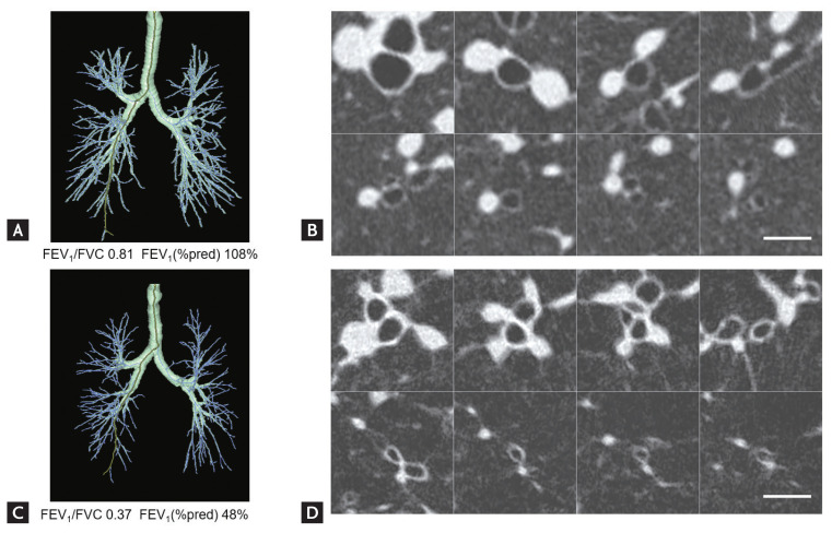
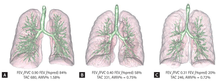
Chronic obstructive pulmonary disease (COPD) is a complex lung disease characterized by a combination of airway disease and emphysema. Emphysema is classified as centrilobular emphysema (CLE), paraseptal emphysema (PSE), or panlobular emphysema (PLE), and airway disease extends from the respiratory, terminal, and preterminal bronchioles to the central segmental airways. Although clinical computed tomography (CT) cannot be used to visualize the small airways, micro-CT has shown that terminal bronchiole disease is more severe in CLE than in PSE and PLE, and micro-CT findings suggest that the loss and luminal narrowing of terminal bronchioles is an early pathological change in CLE. Furthermore, the introduction of ultra-high-resolution CT has enabled direct evaluation of the proximal small (1 to 2-mm diameter) airways, and new CT analytical methods have enabled estimation of small airway disease and prediction of future COPD onset and lung function decline in smokers with and without COPD. This review discusses the literature on micro-CT and the technical advancements in clinical CT analysis for COPD. Hopefully, novel micro-CT findings will improve our understanding of the distinct pathogeneses of the emphysema subtypes to enable exploration of new therapeutic targets, and sophisticated CT imaging methods will be integrated into clinical practice to achieve more personalized management.
Keywords: Airway obstruction; Emphysema; Pathology; Pulmonary disease, chronic obstructive; Tomography, X-ray computed.
Conflict of interest statement
Conflict of interest
This work was partially supported by a grant from the Fujifilm Corporation. No potential conflict of interest relevant to this article was reported.
Figures
References
-
- Singh D, Agusti A, Anzueto A, et al. Global strategy for the diagnosis, management, and prevention of chronic obstructive lung disease: the GOLD science committee report 2019. Eur Respir J. 2019;53:1900164. - PubMed
-
- Gevenois PA, De Vuyst P, de Maertelaer V, et al. Comparison of computed density and microscopic morphometry in pulmonary emphysema. Am J Respir Crit Care Med. 1996;154:187–192. - PubMed
-
- Grydeland TB, Dirksen A, Coxson HO, et al. Quantitative computed tomography measures of emphysema and airway wall thickness are related to respiratory symptoms. Am J Respir Crit Care Med. 2010;181:353–359. - PubMed