. 2021 Oct 26;12(5):e0205721.
doi: 10.1128/mBio.02057-21. Epub 2021 Oct 5.

Revisiting the Role of Toxoplasma gondii ERK7 in the Maintenance and Stability of the Apical Complex

Affiliations

Revisiting the Role of Toxoplasma gondii ERK7 in the Maintenance and Stability of the Apical Complex

Nicolas Dos Santos Pacheco et al. mBio. .

Abstract

Toxoplasma gondii extracellular signal-regulated kinase 7 (ERK7) is known to contribute to the integrity of the apical complex and to participate in the final step of conoid biogenesis. In the absence of ERK7, mature parasites lose their conoid complex and are unable to glide, invade, or egress from host cells. In contrast to a previous report, we show here that the depletion of ERK7 phenocopies the depletion of the apical cap protein AC9 or AC10. The absence of ERK7 leads to the loss of the apical polar ring (APR), the disorganization of the basket of subpellicular microtubules (SPMTs), and a severe impairment in microneme secretion. Ultrastructure expansion microscopy (U-ExM), coupled to N-hydroxysuccinimide ester (NHS-ester) staining on intracellular parasites, offers an unprecedented level of resolution and highlights the disorganization of the rhoptries as well as the dilated plasma membrane at the apical pole in the absence of ERK7. Comparative proteomics analysis of wild-type and ERK7-depleted parasites confirmed the disappearance of known apical complex proteins, including markers of the apical polar ring and a new apical cap named AC11. Concomitantly, the absence of ERK7 led to an accumulation of microneme proteins, resulting from the defect in the exocytosis of the organelles. AC9-depleted parasites were included as controls and exhibited an increase in inner membrane complex proteins, with two new proteins assigned to this compartment, namely, IMC33 and IMC34. IMPORTANCE The conoid is an enigmatic, dynamic organelle positioned at the apical tip of the coccidian subgroup of the Apicomplexa, close to the apical polar ring (APR) from which the subpellicular microtubules (SPMTs) emerge and through which the secretory organelles (micronemes and rhoptries) reach the plasma membrane for exocytosis. In Toxoplasma gondii, the conoid protrudes concomitantly with microneme secretion, during egress, motility, and invasion. The conditional depletion of the apical cap structural protein AC9 or AC10 leads to a disorganization of SPMTs as well as the loss of the APR and conoid, resulting in a microneme secretion defect and a block in motility, invasion, and egress. We show here that the depletion of the kinase ERK7 phenocopies AC9 and AC10 mutants. The combination of ultrastructure expansion microscopy and NHS-ester staining revealed that ERK7-depleted parasites exhibit a dilated apical plasma membrane and an altered positioning of the rhoptries, while electron microscopy images unambiguously highlight the loss of the APR.

Keywords: Apicomplexa; Toxoplasma gondii; apical cap; apicomplexan parasites; comparative proteomics; conoid; egress; extracellular signal-regulated kinase; host cell invasion; invasion; microneme secretion; microtubule; motility; subpellicular microtubules.


Figures

FIG 1
FIG 1
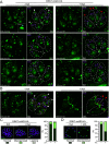
ERK7 depletion caused major cytoskeletal defects at the apical pole. (A) Extracellular parasites were extracted with deoxycholate (DOC) and placed on gelatin-coated coverslips. Depletion of ERK7 caused the collapse of the microtubular cytoskeleton as only single microtubules can be visualized by IFAs with anti-acetylated tubulin (AcTubulin) antibody. Bars = 2 μm. (B) Ultrastructure expansion microscopy (U-ExM) highlighted the absence of the APR and consequently an enlargement of the apical pole and disorganization of the cytoskeleton in ERK7-depleted parasites. On the right are quantifications of abnormal parasites (missing a conoid and/or with an enlarged apical pole and/or disorganized microtubules) versus normal parasites by U-ExM. For the four conditions presented, 200 parasites were counted for each of the three independent biological replicates. Bars = 2 μm. ****, P < 0.0001. (C) U-ExM was applied under intracellular conditions with AC2 tagged in the ERK7-inducible strain. Upon the addition of IAA, the conoid is missing exclusively in the mother cell, and in some severe cases of SPMT disorganization, AC2 staining disappears from the apical cap as well (arrowhead). Bars = 2 μm.
FIG 2
FIG 2
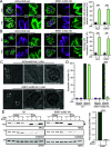
Loss of APR and APR markers in ERK7-depleted parasites. (A) RNG1, a late marker of parasite division, failed to be incorporated into most of the parasite APRs. An RNG1 signal could be detected in the parasite cytoplasm/residual body (asterisk). Western blot analysis confirmed that RNG1 is not degraded. Bars = 2 μm. (B and C) Two additional APR markers, APR1 and KinA, are incorporated early during daughter cell formation and are exclusively lost in mature parasites following the depletion of ERK7. Western blot analysis of extracellular parasites treated with IAA showed that both proteins are degraded. Arrowheads indicate the mother cell apex. Bars = 2 μm. (D) Quantification of vacuoles in which a parasite displays a normal apical RNG1, APR1, or KinA marker. For the six conditions presented, 200 vacuoles were counted for each of the three independent biological replicates. ****, P < 0.0001. (E) EM pictures showing that ERK7 depletion caused the physical loss of the APR and an enlargement of the apical pole of extracellular parasites, in good agreement with the IFA using the RNG1, APR1, and KinA markers. White arrowheads indicate a normal APR; black arrowheads indicate a missing APR. Bars = 200 nm.
FIG 3
FIG 3
NHS-ester coupled with U-ExM highlights conoid loss, rhoptry disorganization, and plasma membrane slackness. (A) Maximum-projection pictures of ERK7-untreated and -treated parasites. The top raw images are extracted from Movies S1 and S3 in the supplemental material (sometimes rotated to have the majority of the parasite apical pole facing up). All images are presented with NHS-ester staining only (left) and with RON9 immunostaining and some annotations (right). In the top raw images, white arrowheads indicate mother cell conoids (light blue arrowhead, intraconoidal microtubules), and red arrowheads indicate the deformation of the apical plasma membrane and the absence of the mother cell conoid. Dotted lines follow the shape of mature parasites (white). Asterisks indicate the basal pole for each vacuole. (B) Same as panel A, with parasites in division (from Movies S2 and S4). Yellow dotted lines follow the shape of the forming daughter cells. (C and D) Regular immunofluorescence highlights rhoptry disorganization using RON9 and ARO antibodies. Quantification of ∼100 vacuoles on a single biological replicate is shown solely for information purposes. Bars = 2 μm.
FIG 4
FIG 4
ERK7-depleted parasites are defective in PM integrity and microneme secretion. (A and B) For both AC9 and ERK7 depletion, some parasites per vacuole showed a defect in plasma membrane integrity at the apical tip (A) and leaking of micronemes (B), highlighted by SAG1 and MIC4 staining, respectively. For the four conditions presented, 200 vacuoles were counted for each of the three independent biological replicates. Bars = 2μm. ***, P < 0.001. (C) Large bright-field images of AC9 and EKR7 parasites grown for 48 h in the presence of IAA. Depleted parasites showed no defects in intracellular growth (intracellular); however, parasites remained trapped inside the floating host cell remnant (detached host cells), suggesting microneme secretion impairment. (D) ERK7 displayed a severe defect in a “standard” induced-egress assay. For each condition presented, 200 vacuoles or PVM remnants were counted for each of three independent biological replicates. ****, P < 0.0001. DMSO, dimethyl sulfoxide. (E) Depletion of ERK7 caused a dramatic block in microneme secretion when the parasites were stimulated with ethanol (EtOH). Anti-MIC2 antibodies were used for secretion (white arrow, full-length MIC2; black arrow, secreted MIC2), anti-catalase (CAT) to assess parasite lysis, and anti-dense granule 1 (GRA1) for constitutive secretion; both pellets and supernatants (ESA) were analyzed. For each of the three independent biological replicates, the intensity of the ESA bands was assessed by band densitometry, and subsequent MIC2/GRA1 ratios are presented (no-IAA ratios are normalized). ****, P < 0.0001.
FIG 5
FIG 5
Comparative proteomics of ERK7- and AC9-depleted parasites. (A and B) Volcano plots showing all the proteins found in ERK7 (A) and AC9 (B) mAID parasites and their differential abundances in auxin-treated parasites versus untreated parasites (LFDR, local false discovery rate). The most significant changes are colored and grouped based on their known and predicted (LOPIT) localizations. On the left (in green) are proteins that are depleted and that majorly localize to the apical complex. The proteins on the right (in magenta) are the ones that are enriched and are found majorly within the micronemes. (C) Venn diagrams showing the significant changes in ERK7 and AC9 (proteins individually depleted or enriched) and the common proteins depleted or enriched in both ERK7 and AC9 auxin-treated parasites. (D) Predicted localization of enriched and depleted proteins in both ERK7 and AC9 according to hyperLOPIT.
FIG 6
FIG 6
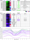
Statistically significant proteins depleted/enriched in both strains. (A and B) Heat maps showing the most significant changes (FDR < 0.05) under both ERK7 and AC9 (auxin-treated) conditions. In green (A) are the proteins that are significantly depleted, and in magenta (B) are the proteins that are significantly enriched in both ERK7 and AC9. Predicted (LOPIT) localizations and previously localized proteins are also indicated (1, 7, 8, 23, 24, 31, 40–47, 51). (C) Expression profile of proteins with no “experimental localization” presented in panel B. Out of 17 proteins, 7 of them (thick lines/shades of magenta) share a similar expression profile with a typical IMC-related protein, namely, IMC1 (cyan). The 10 others, with different expression profiles, are presented with thin gray lines. Data were retrieved from the ToxoDB website [Transcriptomic data “T. gondii ME49 Cell Cycle Expression Profiles (RH)” from reference 52].
FIG 7
FIG 7
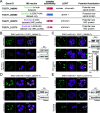
Validation of potential new apical and IMC proteins. (A) Table presenting the four candidates successfully tagged in the ERK7-mAID strain. (B) TGGT1_246040 localizes as discrete dots throughout the cytoplasm of nondividing and dividing parasites. Bars = 2 μm. (C) AC11 (TGGT1_266080) localizes only at the apical cap of mature cells and is affected by the disorganization of the apical pole in ERK7-depleted parasites. Bars = 2 μm. The graph presents the quantification of vacuoles with at least one parasite displaying abnormal AC11 staining. Around 200 vacuoles were counted for each of the three independent biological replicates. (D) IMC33 (TGGT1_282070) decorates the IMC of forming daughter cells. It appears early during daughter cell biogenesis and seems to accumulate at the growing end of the daughter cells before their emergence. Bars = 2 μm. (E) IMC34 (TGGT1_212770) decorates the IMC of forming daughter cells only. The black arrowhead indicates nondividing parasites with no IMC34 signal, while the white arrowhead indicates dividing parasites displaying clear IMC34 staining. Bars = 2 μm.

Similar articles

Cited by

References

    1. Tosetti N, Dos Santos Pacheco N, Bertiaux E, Maco B, Bournonville L, Hamel V, Guichard P, Soldati-Favre D. 2020. Essential function of the alveolin network in the subpellicular microtubules and conoid assembly in Toxoplasma gondii. Elife 9:e56635. doi:10.7554/eLife.56635. - DOI - PMC - PubMed
    1. Back PS, O’Shaughnessy WJ, Moon AS, Dewangan PS, Hu X, Sha J, Wohlschlegel JA, Bradley PJ, Reese ML. 2020. Ancient MAPK ERK7 is regulated by an unusual inhibitory scaffold required for Toxoplasma apical complex biogenesis. Proc Natl Acad Sci USA 117:12164–12173. doi:10.1073/pnas.1921245117. - DOI - PMC - PubMed
    1. O’Shaughnessy WJ, Hu X, Beraki T, McDougal M, Reese ML. 2020. Loss of a conserved MAPK causes catastrophic failure in assembly of a specialized cilium-like structure in Toxoplasma gondii. Mol Biol Cell 31:881–888. doi:10.1091/mbc.E19-11-0607. - DOI - PMC - PubMed
    1. Hu K, Roos DS, Murray JM. 2002. A novel polymer of tubulin forms the conoid of Toxoplasma gondii. J Cell Biol 156:1039–1050. doi:10.1083/jcb.200112086. - DOI - PMC - PubMed
    1. Dos Santos Pacheco N, Tosetti N, Koreny L, Waller RF, Soldati-Favre D. 2020. Evolution, composition, assembly, and function of the conoid in Apicomplexa. Trends Parasitol 36:688–704. doi:10.1016/j.pt.2020.05.001. - DOI - PubMed

Publication types

MeSH terms

Substances

LinkOut - more resources