

Two target genes based multiple cross displacement amplification combined with a lateral flow biosensor for the detection of Mycobacterium tuberculosis complex
- PMID: 34607556
- PMCID: PMC8491432
- DOI: 10.1186/s12866-021-02328-6
Two target genes based multiple cross displacement amplification combined with a lateral flow biosensor for the detection of Mycobacterium tuberculosis complex
Abstract
Background: Tuberculosis (TB) is a serious chronic infectious disease caused by Mycobacterium tuberculosis complex (MTBC). Hence, the development of a novel, simple, rapid and sensitive method to detect MTBC is of great significance for the prevention and treatment of TB.
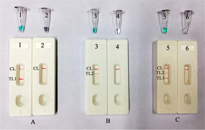
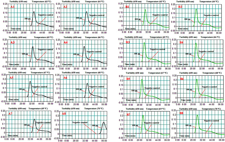
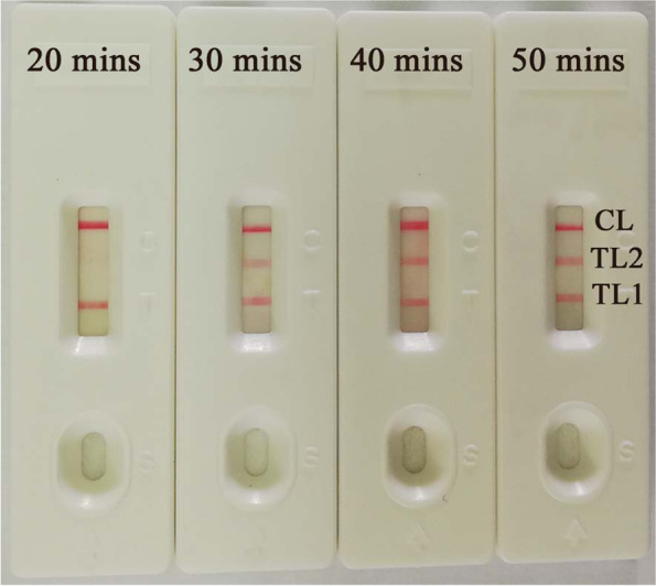
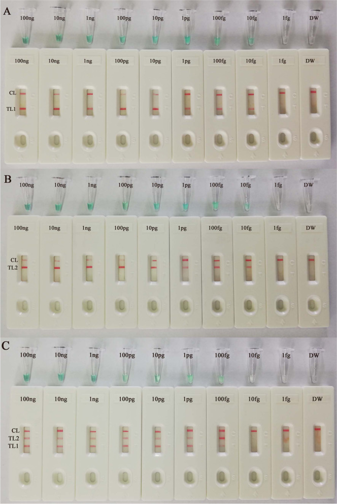
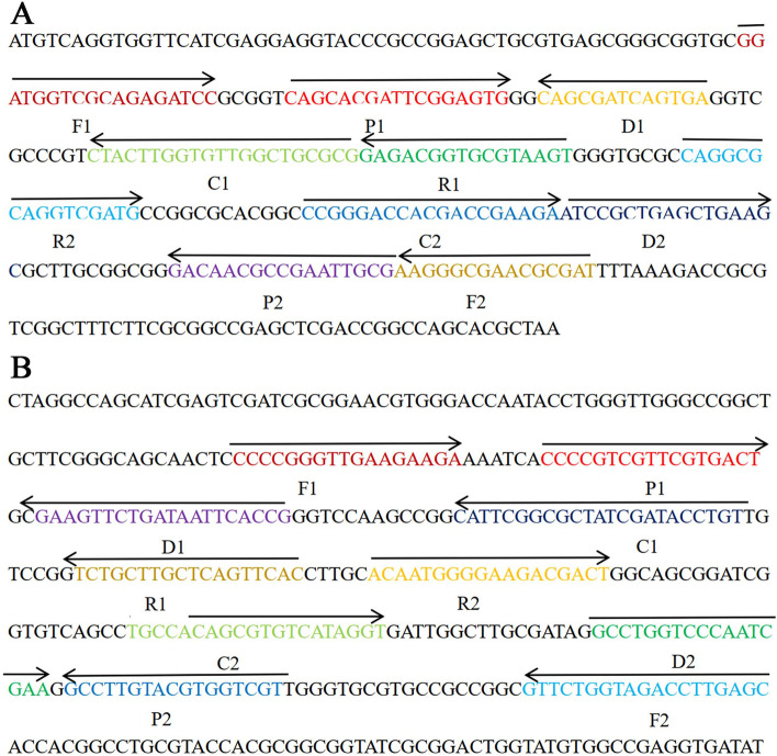
Results: In this study, multiple cross displacement amplification (MCDA) combined with a nanoparticle-based lateral flow biosensor (LFB) was developed to simultaneously detect two target genes (IS6110 and mpb64) of MTBC (MCDA-LFB). One suite of specific MCDA primers designed for the IS6110 and mpb64 genes was validated using genomic DNA extracted from the reference strain H37Rv. The MCDA amplicons were analyzed using a real-time turbidimeter, colorimetric indicator (malachite green, MG) and LFBs. The optimal amplification temperature and time were confirmed, and the MCDA-LFB method established in the current report was evaluated by detecting various pathogens (i.e., reference strains, isolates and clinical sputum samples). The results showed that the two sets of MCDA primers targeting the IS6110 and mpb64 genes could effectively detect MTBC strains. The optimal reaction conditions for the MCDA assay were determined to be 67 °C for 35 min. The MCDA assay limit of detection (LoD) was 100 fg per reaction for pure genomic DNA. The specificity of the MCDA-LFB assay was 100%, and there were no cross-reactions for non-MTBC strains. For sputum samples and MTBC strain detection, the positive rate of MCDA-LFB for the detection of MTBC strains was consistent with seminested automatic real-time PCR (Xpert MTB/RIF) and higher than acid-fast staining (AFS) and culture assays when used for sputum samples. The MCDA-LFB assay was a rapid tool, and the whole procedure for MCDA-LFB, including DNA template preparation, MCDA reaction and amplification product analysis, was completed within 70 min.
Conclusion: The MCDA-LFB assay targeting the IS6110 and mpb64 genes is a simple, rapid, sensitive and reliable detection method, and it has potential significance for the prevention and treatment of TB.
Keywords: Detection; Diagnosis; Lateral flow biosensor; Multiple cross displacement amplification; Mycobacterium tuberculosis complex.
© 2021. The Author(s).
Conflict of interest statement
All of the authors declare that there are no competing interests in this article.
Figures
Similar articles
-
Establishment and Application of a Multiple Cross Displacement Amplification Coupled With Nanoparticle-Based Lateral Flow Biosensor Assay for Detection of Mycoplasma pneumoniae.Front Cell Infect Microbiol. 2019 Sep 23;9:325. doi: 10.3389/fcimb.2019.00325. eCollection 2019. Front Cell Infect Microbiol. 2019. PMID: 31608243 Free PMC article.
-
Development and Preliminary Application of Multiplex Loop-Mediated Isothermal Amplification Coupled With Lateral Flow Biosensor for Detection of Mycobacterium tuberculosis Complex.Front Cell Infect Microbiol. 2021 Apr 27;11:666492. doi: 10.3389/fcimb.2021.666492. eCollection 2021. Front Cell Infect Microbiol. 2021. PMID: 33987108 Free PMC article.
-
Development and Clinical Validation of Multiple Cross Displacement Amplification Combined With Nanoparticles-Based Biosensor for Detection of Mycobacterium tuberculosis: Preliminary Results.Front Microbiol. 2019 Sep 13;10:2135. doi: 10.3389/fmicb.2019.02135. eCollection 2019. Front Microbiol. 2019. PMID: 31572340 Free PMC article.
-
Loop-Mediated Isothermal Amplification Coupled With Nanoparticle-Based Lateral Biosensor for Rapid, Sensitive, and Specific Detection of Bordetella pertussis.Front Bioeng Biotechnol. 2022 Feb 8;9:797957. doi: 10.3389/fbioe.2021.797957. eCollection 2021. Front Bioeng Biotechnol. 2022. PMID: 35211469 Free PMC article. Review.
-
Lateral flow biosensor combined with loop-mediated isothermal amplification for simple, rapid, sensitive, and reliable detection of Brucella spp.Infect Drug Resist. 2019 Jul 30;12:2343-2353. doi: 10.2147/IDR.S211644. eCollection 2019. Infect Drug Resist. 2019. PMID: 31440069 Free PMC article. Review.
Cited by
-
Multiple cross displacement amplification combined with nanoparticle-based lateral flow biosensor for rapid and sensitive detection of Epstein-Barr virus.Front Cell Infect Microbiol. 2024 Jan 8;13:1321394. doi: 10.3389/fcimb.2023.1321394. eCollection 2023. Front Cell Infect Microbiol. 2024. PMID: 38259964 Free PMC article.
-
Development and preliminary assessment of a CRISPR-Cas12a-based multiplex detection of Mycobacterium tuberculosis complex.Front Bioeng Biotechnol. 2023 Aug 25;11:1233353. doi: 10.3389/fbioe.2023.1233353. eCollection 2023. Front Bioeng Biotechnol. 2023. PMID: 37711452 Free PMC article.
-
Development of CRISPR/Cas12b-Based Multiple Cross Displacement Amplification Technique for the Detection of Mycobacterium tuberculosis Complex in Clinical Settings.Microbiol Spectr. 2023 Mar 28;11(2):e0347522. doi: 10.1128/spectrum.03475-22. Online ahead of print. Microbiol Spectr. 2023. PMID: 36975805 Free PMC article.
-
Nanoparticle-based biosensor integrated with multiple cross-displacement amplification for visual and rapid identification of hepatitis B virus and hepatitis C virus.Microbiol Spectr. 2025 May 6;13(5):e0173824. doi: 10.1128/spectrum.01738-24. Epub 2025 Apr 15. Microbiol Spectr. 2025. PMID: 40231683 Free PMC article.
-
Development and clinical evaluation of a real-time multiple cross displacement amplification assay for rapid and sensitive detection of Mycobacterium tuberculosis.Heliyon. 2024 May 24;10(11):e31901. doi: 10.1016/j.heliyon.2024.e31901. eCollection 2024 Jun 15. Heliyon. 2024. PMID: 38845879 Free PMC article.
References
-
- Cadmus SI, Jenkins AO, Godfroid J, Osinusi K, Adewole IF, Murphy RL, Taiwo BO. Mycobacterium tuberculosis and Mycobacterium africanum in stools from children attending an immunization clinic in Ibadan, Nigeria. Int J Infect Dis. 2009;13(6):740–744. doi: 10.1016/j.ijid.2008.11.016. - DOI - PMC - PubMed
-
- Organization WHO: Global tuberculosis report 2019. Online at https://www.who.int/teams/global-tuberculosis-programme/tb-reports/globa....
Publication types
MeSH terms
Substances
LinkOut - more resources
Full Text Sources
Medical
Molecular Biology Databases
