Review
. 2022 Jan;47(1):82-97.
doi: 10.1016/j.tibs.2021.09.001. Epub 2021 Oct 2.

Canary in a coal mine: collided ribosomes as sensors of cellular conditions

Affiliations
Review

Canary in a coal mine: collided ribosomes as sensors of cellular conditions

Kyusik Q Kim et al. Trends Biochem Sci. 2022 Jan.

Abstract

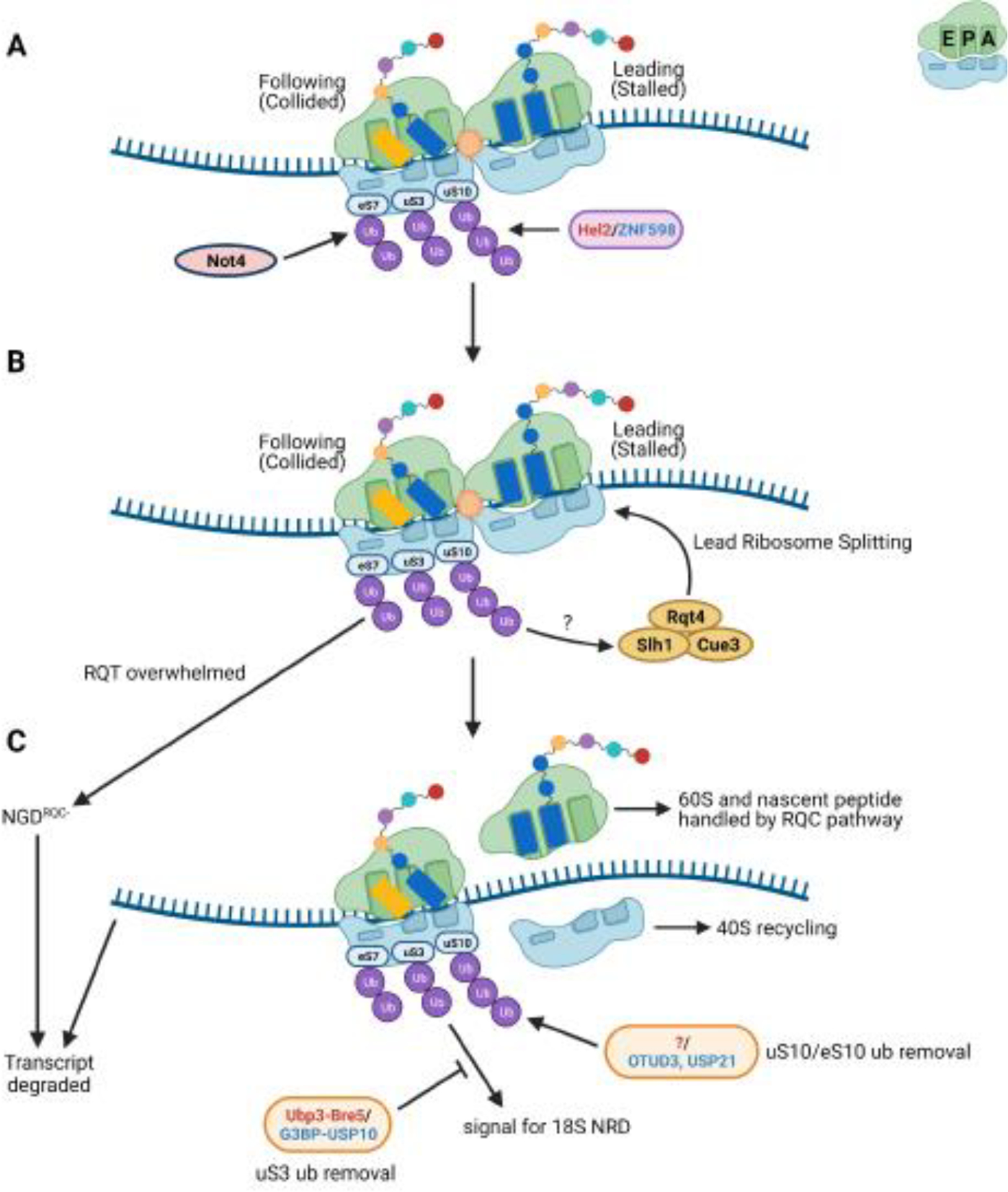
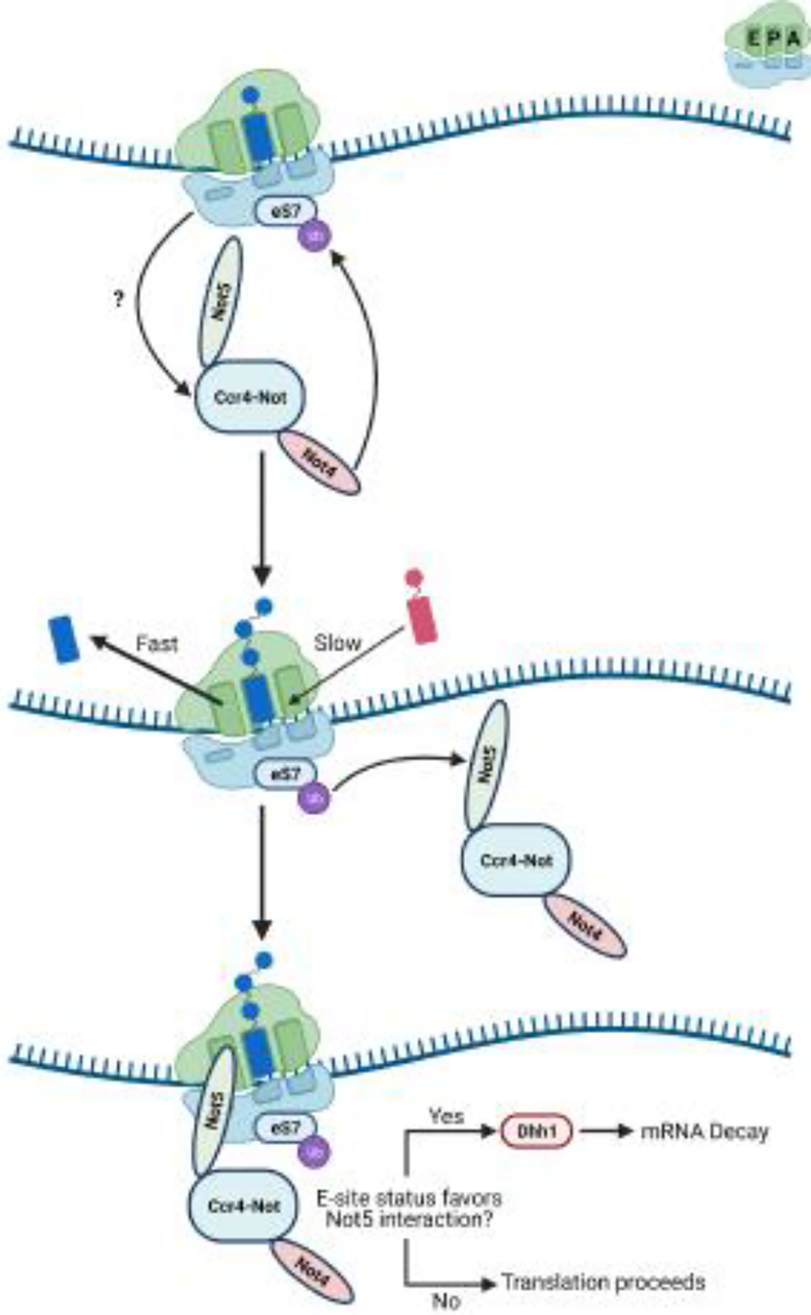
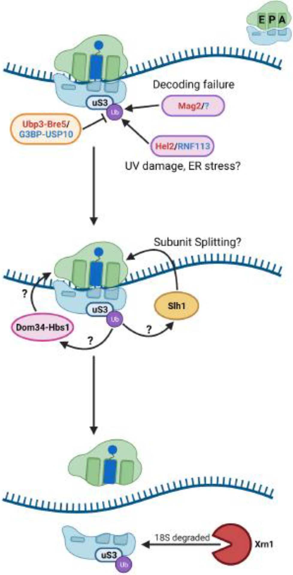
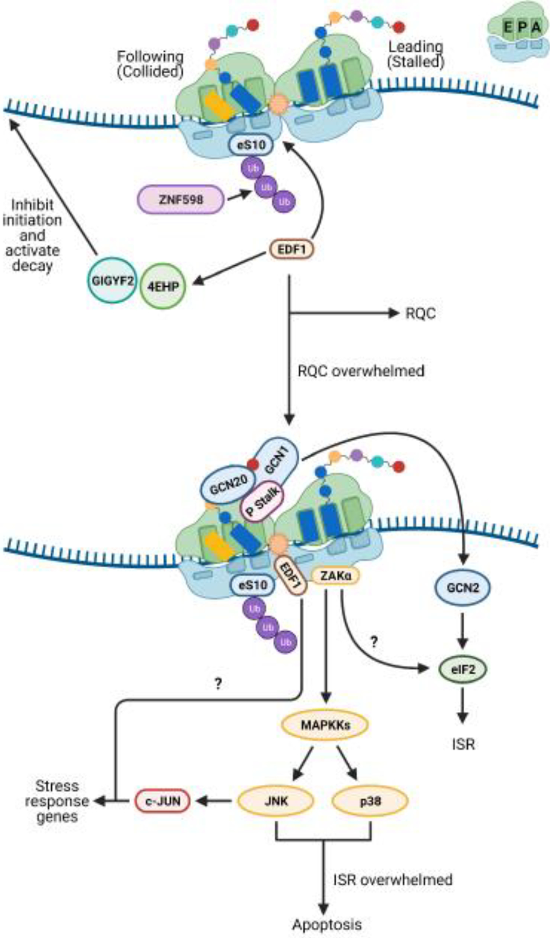
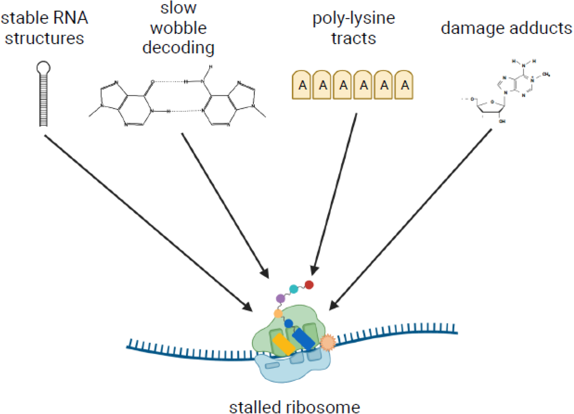
The recent discovery that collision of ribosomes triggers quality control and stress responses in eukaryotes has shifted the perspective of the field. Collided eukaryotic ribosomes adopt a unique structure, acting as a ubiquitin signaling platform for various response factors. While several of the signals that determine which downstream pathways are activated have been uncovered, we are only beginning to learn how the specificity for the activation of each process is achieved during collisions. This review will summarize those findings and how ribosome-associated factors act as molecular sentinels, linking aberrations in translation to the overall cellular state. Insights into how cells respond to ribosome collision events will provide greater understanding of the role of the ribosome in the maintenance of cellular homeostasis.

Keywords: collisions; integrated stress response; mRNA surveillance; quality control; ribosome; signaling.


Conflict of interest statement

Declaration of interests None are declared.

Figures

Figure 1.
Figure 1.. Signaling on collided ribosomes and its impact on ribosome fate.
Small cartoon depicting the A, P, and E sites is in the top right of the figure. (A) Ubiquitination of ribosomal proteins uS10 and uS3 by Hel2 in yeast, and eS10, uS10, and uS3 by ZNF598 in mammals, occurs on stable collided ribosome structures. In yeast, eS7 is ubiquitinated by Not4 via an unknown mechanism and polyubiquitinated by Hel2 during stalling. Which ribosomes within the collision units are ubiquitinated remains unclear. Given that Hel2/ZNF598 has been proposed to recognize the rotated state of collided ribosomes, one potential model is that only the collided ribosomes are ubiquitinated, enabling distinction of the stalled ribosome. However, uS3 and eS7 may show different signaling patterns. For simplicity, only the collided ribosome is depicted as ubiquitinated. (B) The stalled ribosome is split by the ribosome-quality control trigger (RQT) complex but the purpose of the ubiquitin marks in this process remains unknown. In the event the RQT complex is overwhelmed, polyubiquitinated eS7 acts as a signal for a secondary mRNA decay mechanism termed NGDRQC-. (C) Once split, the nascent peptide-bound 60S is handled by the RQC pathway and the 40S is presumably returned to the free 40S pool. The transcript is degraded by downstream mRNA decay pathways. eS10 and uS10 ubiquitin marks can be removed by the mammalian de-ubiquitinases OTUD3 and USP21. The homologous yeast deubiquitinase is yet to be identified (denoted by “?” in the figure). A yet undetermined uS3 ubiquitin signal triggers 18S NRD but can be opposed by the action of the de-ubiquitination complexes Ubp3-Bre5 in yeast or G3BP-USP10 in mammals. Names of the factors in red/blue denote the yeast and mammalian factor, respectively.
Figure 2.
Figure 2.. Model for Not5-mediated mRNA decay on the ribosome.
Small cartoon depicting the A, P, and E sites is in the top right of the figure. In yeast, Not5 is anchored to the ribosome via Not4-mediated eS7 ubiquitination. When and how Not4 targets ribosome for eS7 ubiquitination is not completely understood. Once on the ribosome, Not5 monitors E-site occupancy as a proxy for A-site decoding efficiency. When E-site occupancy is low, Not5 recruits the DEAD-box helicase Dhh1 to initiate mRNA decay.
Figure 3.
Figure 3.. Nonfunctional ribosome decay (NRD) of 18S rRNA.
Small cartoon depicting the A, P, and E sites is in the top right of the figure. In yeast, uS3 ubiquitination by Mag2 in response to decoding failure by the small subunit leads to subunit splitting and degradation of the 18S rRNA. uS3 can be ubiquitinated in response to other stimuli, such as UV damage, and deubiquitinated by Ubp3-Bre5 (yeast) or G3BP-USP10 (human). How these signaling processes are coordinated remains unclear. The role of ribosome splitting factors Slh1 and Dom34-Hbs1, as well as their interplay, during 18S NRD in yeast also remain undetermined. Names of the factors in red/blue denote the yeast and mammalian factor, respectively.
Figure 4.
Figure 4.. Collided ribosomes are a signaling hub for stress response factors.
Small cartoon depicting the A, P, and E sites is in the top right of the figure. In mammalian cells, when numbers of collided ribosomes remain low, the ribosome-associated quality control (RQC) pathway is able to deal with stalled ribosomes ubiquitinated by ZNF598. The factor EDF1 is recruited to the small subunit of the collided ribosome by a still-undetermined mechanism and potentially acts to prevent frameshifting during stall resolution. EDF1 also appears to play a role in the recruitment of the factors GIGYF2 and 4EHP to repress initiation on aberrant transcripts. When collision frequencies become elevated, such as during nutrient stress, the RQC machinery is overwhelmed, and alternative response pathways become activated. The P stalk of long-lived collided ribosome species may activate the integrated stress response (ISR) kinase GCN2. It is still unknown how GCN2’s cofactors GCN1 and GCN20 coordinate with the P stalk to activate GCN2. Long-lived collisions may also activate the mitogen activated kinase kinase kinase (MAPKKK) ZAKα. The mechanism by which ZAKα is activated has not been clearly determined. ZAKα signaling activates p38 and c-Jun N-terminal kinase (JNK) and might play a role in ISR signaling. JNK signaling leads to upregulation of the stress response transcription factor c-Jun, which in turn upregulates stress response genes. In addition to its other activities, EDF1 appears to contribute to c-Jun transcriptional activity via an unknown mechanism. In the event the ISR fails to restore homeostasis, p38 and JNK signaling leads to apoptosis.
None
None

References

    1. García-Muse T and Aguilera A (2016) Transcription-replication conflicts: How they occur and how they are resolved. Nat. Rev. Mol. Cell Biol 17, 553–563 - PubMed
    1. Sørensen MA and Pedersen S (1991) Absolute in vivo translation rates of individual codons in Escherichia coli. The two glutamic acid codons GAA and GAG are translated with a threefold difference in rate. J. Mol. Biol 222, 265–280 - PubMed
    1. Mitarai N et al. (2008) Ribosome Collisions and Translation Efficiency: Optimization by Codon Usage and mRNA Destabilization. J. Mol. Biol 382, 236–245 - PubMed
    1. Farabaugh PJ (1996) Programmed translational frameshifting. Annu. Rev. Genet 30, 507–528 - PubMed
    1. Richter JD and Coller J (2015) Pausing on Polyribosomes: Make Way for Elongation in Translational Control. Cell 163, 292–300 - PMC - PubMed

Publication types

MeSH terms

LinkOut - more resources