Review
. 2022 Feb;32(2):151-164.
doi: 10.1016/j.tcb.2021.09.002. Epub 2021 Oct 1.

Metabolic and epigenetic regulation of endoderm differentiation

Affiliations
Review

Metabolic and epigenetic regulation of endoderm differentiation

Yi Fang et al. Trends Cell Biol. 2022 Feb.

Abstract

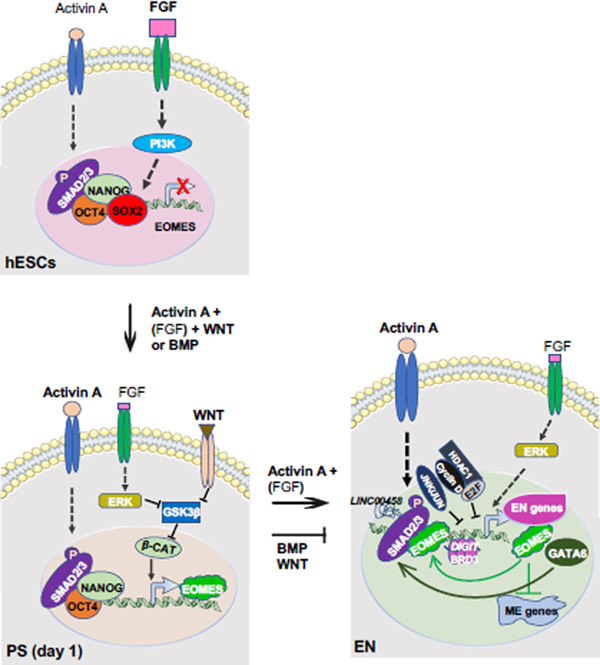
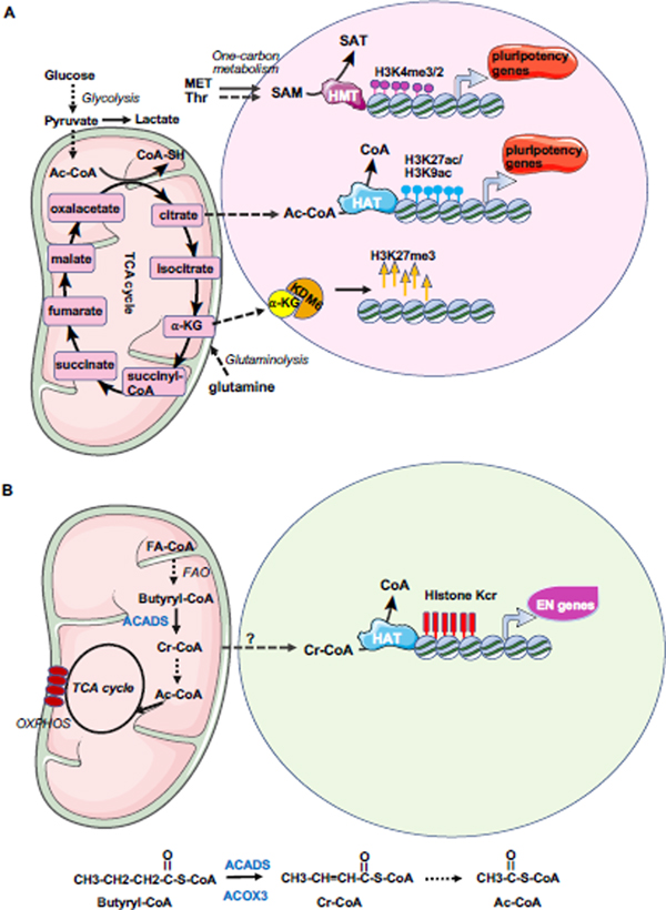
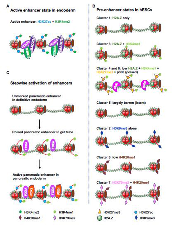
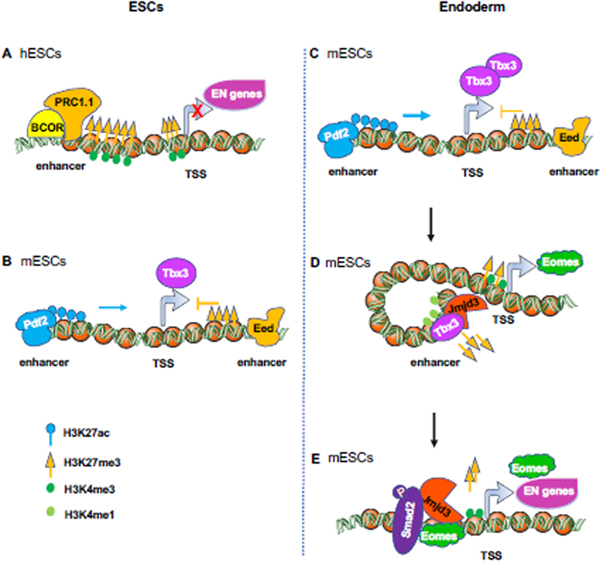
The endoderm, one of the three primary germ layers, gives rise to lung, liver, stomach, intestine, colon, pancreas, bladder, and thyroid. These endoderm-originated organs are subject to many life-threatening diseases. However, primary cells/tissues from endodermal organs are often difficult to grow in vitro. Human pluripotent stem cells (hPSCs), therefore, hold great promise for generating endodermal cells and their derivatives for the development of new therapeutics against these human diseases. Although a wealth of research has provided crucial information on the mechanisms underlying endoderm differentiation from hPSCs, increasing evidence has shown that metabolism, in connection with epigenetics, actively regulates endoderm differentiation in addition to the conventional endoderm inducing signals. Here we review recent advances in metabolic and epigenetic regulation of endoderm differentiation.

Keywords: endoderm differentiation; endodermal gene expression; epigenetic remodeling; histone crotonylation; metabolic switch.


Conflict of interest statement

Declaration of interests The authors declare no competing interests.

Figures

Figure 1.
Figure 1.. Canonical developmental signals and factors important for differentiation of PS and endoderm.
In primed hESCs, low Activin A and high FGF signaling promote the expression of pluripotency genes via their downstream effectors like SMAD2/3 and PI3K, respectively. OCT4, SOX2, and NANOG bind to the regulatory elements of EOMES and repress its expression in undifferentiated hESCs. After induction of endoderm differentiation by exogeneous Activin A with BMP or WNT, reduction of SOX2 allows NANOG to promote EOMES expression. Meanwhile, low level of FGF signaling activates ERK (MAPK), which inhibits GSK3β, an inhibitor of WNT/β-catenin signaling. Exogeneous WNT signals also inhibit GSK3β to activate β-catenin to promote EOMES expression. EOMES then interacts with phosphorylated SMAD2/3 to induce itself, GATA6, and genes characteristically expressed in the PS and endoderm. After PS is generated, exogeneous Nodal/Activin A and endogenous FGF [(FGF)] signaling effectively differentiate cells into endoderm in the following days, while BMP and WNT signaling promotes PS to mesoderm lineage and represses endoderm differentiation. Additionally, GATA6, LINC00458 which interacts with SMAD2/3, and BRD3 condensates with DIGIT also promote endoderm differentiation. Cyclin D which recruits E2F and HDAC1, and JNK/JUN family bind to endoderm loci to block endoderm differentiation. (FGF): endogenous FGF; β-CAT: β-catenin, EN: endoderm; ME: mesoderm.
Figure 2.
Figure 2.. Metabolic influence on ESC maintenance and endoderm differentiation.
(A) hESCs heavily rely on glycolysis and acetyl-CoA, and one-carbon metabolism of methionine and SAM for maintenance of epigenetics required for pluripotency. mESCs are dependent on glutamine metabolism and α-KG as well as one-carbon metabolism of threonine and SAM for maintaining pluripotency and the epigenetic status. Ac-CoA: acetyl-CoA; HMT: histone methyltransferase; HAT: histone acetyltransferase, SAM: S-adenosylmethionine, α-KG: α-ketoglutarate. (B) Crotonyl-CoA from fatty acid oxidation (FAO) enhances histone crotonylation on endodermal genes and promotes endoderm differentiation. Upon endoderm differentiation, butyryl-CoA, a short-chain fatty acyl-CoA which may be produced from the longer-chain fatty-acyl-CoA via β-oxidation (or fatty acid oxidation), is catalyzed to produce crotonyl-CoA by ACADS. Crotonyl-CoA can be degraded to form 2 molecules of acetyl-CoA, which enters TCA cycle to reduce NAD+ to NADH for oxidative phosphorylation. Meanwhile, crotonyl-CoA can be present in the nucleus to enhance histone crotonylation on the regulatory elements of endodermal genes to promote endodermal gene expression. Kcr: lysine crotonylation; Cr-CoA: crotonyl-CoA, FAO: fatty acid oxidation; FA-CoA: fatty acyl-CoA; OXPHOS: oxidative phosphorylation; ACADS: acyl-CoA dehydrogenase short chain (in mitochondria); ACOX3: acyl-CoA oxidase (in peroxisome).
Figure 3.
Figure 3.. States of endodermal gene enhancers in hESCs and endodermal cells.
(A) Active enhancers are highly enriched for H3K27ac and H3K4me2, and are co-occupied by SMAD2/3/4, EOMES and FOXH1. The figure is adapted from [8] (B) Chip-seq was performed in undifferentiated hESCs with chromatin marks and modifiers including H3K4me1, H3K4me2, H3K4me3, H2A.Z, H3K27me3, H3K9me3, H3K27ac, H3K9ac, H3K79me2, H4K20me1, CHD1, CHD7, EZH2, HDAC2, RBBP5, JARID1A and p300. The abundance of the marks/modifiers in the future endoderm enhancers were analyzed, organized by unbiased clustering, and illustrated. The active enhancer state in endodermal cells is highly enriched for H3K27ac and H3K4me2. Cluster 4 and 8 are characterized by low H2A.Z, H3K4me1, H3K27me3 and p300, representing the “poised” enhancer state for quick activation. H2A.Z renders nucleosome unstable and facilitates transcriptional factors to bind DNA. Cluster 2 is enriched by H2A.Z and H3K4me1, marking the enhancer for subsequent activation. Those pre-enhancers are devoid of any chromatin marks/modifiers as illustrated in cluster 5, are latent in activation. The presence of multiplicity of pre-enhancer states allows a precise control of enhancer activation under the influence of both lineage inducing signals and effectors. The figure is adapted from [8]. (C) Stepwise activation of pancreatic enhancers. Pancreatic enhancers are unmarked in definitive endoderm, are poised by H3K4me1 and FOXA proteins in gut tube, and are active in pancreatic endoderm after H3K27ac deposition and PDX1 binding. The figure is adapted from [51].
Figure 4.
Figure 4.. Dynamic regulation of H3K27 trimethylation on endodermal genes in ESCs and endodermal cells.
(A) BCOR, a member of PRC1.1 complex, specifically represses the expression of endodermal and mesodermal genes in hESCs through enhancement of H3K27me3. (B-C) Eed and Dpf2 antagonize the expression of Tbx3 in mESCs. (B) Eed, a subunit of PRC2 complex binds an intragenic Tbx3 enhancer to oppose Dpf2 (a subunit of SWI-SNF complex)-dependent Tbx3 expression and mesendodermal differentiation in mESCs. (C) Dpf2 binds the distal enhancer of Tbx3, increases the H3K27ac level, and promotes the expression of Tbx3 during mesendodermal differentiation of mESCs. (D-E) Jmjd3 and Eomes function in a positive feedback loop to promote endoderm differentiation from mESCs. (D) Jmjd3 physically associates with Tbx3, then binds to a bivalent enhancer (marked by H3K4me1 and H3K27me3) of Eomes, which in turn leads to a reduction of H3K27me3 deposition in the enhancer, and an interaction between enhancer and promoter, thereby promoting Eomes expression. (E) Jmjd3 and Smad2, recruited by Eomes, co-bind to the bivalent promoters (marked by H3K4me3 and H3K27me3) of core endoderm differentiation genes such as Eomes, Sox17, Gsc, Gata6, Mixl1 and Foxa2 to remove H3K27me3 for their promoter activation[48].

Similar articles

Cited by

References

    1. Nowotschin S et al. (2019) The endoderm: a divergent cell lineage with many commonalities. Development 146 (11). - PMC - PubMed
    1. Yiangou L et al. (2018) Human Pluripotent Stem Cell-Derived Endoderm for Modeling Development and Clinical Applications. Cell Stem Cell 22 (4), 485–499. - PubMed
    1. Tam PP and Beddington RS (1987) The formation of mesodermal tissues in the mouse embryo during gastrulation and early organogenesis. Development 99 (1), 109–26. - PubMed
    1. Lawson KA et al. (1991) Clonal analysis of epiblast fate during germ layer formation in the mouse embryo. Development 113 (3), 891–911. - PubMed
    1. Tada S et al. (2005) Characterization of mesendoderm: a diverging point of the definitive endoderm and mesoderm in embryonic stem cell differentiation culture. Development 132 (19), 4363–74. - PubMed

Publication types