Review
. 2022 Jul 1;14(7):a040840.
doi: 10.1101/cshperspect.a040840.

Building and Maintaining the Skin

Affiliations
Review

Building and Maintaining the Skin

Ya-Chieh Hsu et al. Cold Spring Harb Perspect Biol. .

Abstract

The skin forms a crucial, dynamic barrier between an animal and the external world. In mammals, three stem cell populations possess robust regenerative potential to maintain and repair the body's protective surface: epidermal stem cells, which maintain the stratified epidermis; hair follicle stem cells, which power the cyclic growth of the hair follicle; and melanocyte stem cells, which regenerate pigment-producing melanocytes to color the skin and hair. These stem cells reside in complex microenvironments ("niches") comprising diverse cellular repertoires that enable stem cells to rejuvenate tissues during homeostasis and regenerate them upon injury. Beyond their niches, skin stem cells can also sense and respond to fluctuations in organismal health or changes outside the body. Here, we review these diverse cellular interactions and highlight how far-reaching signals can be transmitted at the local level to enable skin stem cells to tailor their actions to suit the particular occasion and optimize fitness.


Figures

Figure 1.
Figure 1.
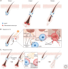
Skin stem cells and their niches. The skin is a complex organ composed of cell types from diverse lineages. With the exception of higher primates, whose eccrine sweat glands and associated stem cells are highly abundant, most mammals have three major skin stem cell populations—epidermal stem cells, hair follicle stem cells, and melanocyte stem cells. These stem cells regenerate in rich microenvironments (niches) replete with diverse cell types, including distinct fibroblast populations, sensory and sympathetic innervations, a vast array of immune cells, blood vessels, lymphatic capillaries, and subcutaneous adipocytes. In addition to their specialized functions, complex interactions among these cell types underlie tissue formation, homeostasis, and repair in the skin. (TAC) Transit-amplifying cell, (DP) dermal papilla.
Figure 2.
Figure 2.
The hair follicle lineage and niche signals that regulate hair follicle stem cells (HFSCs). (A) Overview of the hair cycle. The telogen hair follicle contains HFSCs that are located at the outer bulge layer and the hair germ, as well as the K6 (keratin 6)+ inner bulge layer, which is made up of differentiated progeny of HFSCs. During anagen, the HFSCs proliferate to generate transit-amplifying cells (TACs), which undergo rapid proliferation and expansion to make all the differentiated layers of the anagen hair follicle (see insert)—the three layers of the hair shaft, the three layers of its channel (the inner root sheath [IRS]), and the companion layer sandwiched between ORS (outer root sheath) and IRS. In catagen, most of the hair follicle cells produced in anagen are destroyed or remolded, sparing some ORS cells to make a new bulge and hair germ that fuels the next hair cycle. (B) Niche signals regulate HFSC quiescence and activation. In telogen, signals from the K6+ bulge, mature adipocytes, and TERM2+ macrophages maintain HFSCs in a quiescent state. During the telogen to anagen transition, activating signals become highly up-regulated in the dermal papilla (DP). Signals from sympathetic neurons, Regulatory T cells (Tregs), adipocyte precursors, and macrophages have all been reported to contribute to HFSC activation in the hair germ. In early anagen, Sonic Hedgehog (SHH) secreted from the TACs further activates HFSCs located in the bulge. SHH also modulates activation signals expressed in the DP and promotes adipocyte precursors to make mature adipocytes. HFSCs are normally wrapped around tightly by lymphatic capillaries, a process mediated by HFSC-derived Angiopoietin-like 7 (ANGPTL7). During early anagen, HFSCs transiently switch to expressing Angiopoietin-like 4 (ANGPTL4) and Netrin-4 (NTN4), which promotes a transient dissociation between HFSCs and lymphatic capillaries to promote hair growth. (APM) Arrector pili muscle, (OSM) Oncostatin M, (NE) norepinephrine, (BMP) bone morphogenetic protein.
Figure 3.
Figure 3.
Melanocyte stem cells (McSCs) and their responses to ultraviolet (UV) and stress. (A) Schematic of the melanocyte lineage during the hair cycle. Differentiated melanocytes are made during anagen to color the newly regenerated hair shaft. (B) UV irradiation leads to DNA damage in the epidermis and up-regulation of p53 and POMC (proopiomelanocortin), a precursor peptide that is processed to form β-endorphin, Adrenocorticotropic hormone (ACTH), and α-MSH (α-melanocyte-stimulating hormone). α-MSH binds to MC1R (melanocortin 1 receptor) to promote McSC migration toward the epidermis. MC1R signaling further up-regulates a transcription factor, MITF, which promotes melanocyte differentiation and melanin production. (EpdSC) Epidermal stem cells. (C) Under stress, sympathetic neurons become highly activated, releasing large amounts of norepinephrine (NE), which binds to the β2 adrenergic receptor (ADRB2) on McSCs to drive their hyperproliferation, differentiation, and permanent depletion.
Figure 4.
Figure 4.
The stratified skin epidermis. Epidermal stem cells (EpdSCs) are located at the basal layer of the epidermis. EpdSCs adhere to the extracellular matrix rich basement membrane through interactions between α3β1 and α6β4 integrins with the laminin-5. EpdSCs can both self-renew and generate progeny that are committed to terminal differentiate. In some cases, differentiation is triggered when a stem cell divides parallel to the plane of the basal layer, but then one of its daughters detaches and moves into the suprabasal layer. In other cases, perpendicular divisions can result in one basal cell remaining basally as a stem cell, while another committed daughter cell is displaced into the suprabasal layer. Irrespective, cells committed to terminal differentiation transition through the spinous layer, the granular layer, and eventually into the dead stratum corneum that sheds from the skin surface. Until the granular:stratum corneum transition, each layer expresses different sets of proteins, including different keratin proteins. Secreted proteins such as insulin-like growth factors (IGFs) and ligands for epidermal growth factor receptors (EGFRs) promote EpdSC proliferation, while Notch signaling regulates the differentiation process.

Similar articles

Cited by

References

    1. Adam RC, Yang H, Ge Y, Lien WH, Wang P, Zhao Y, Polak L, Levorse J, Baksh SC, Zheng D, et al. 2018. Temporal layering of signaling effectors drives chromatin remodeling during hair follicle stem cell lineage progression. Cell Stem Cell 22: 398–413.e7. 10.1016/j.stem.2017.12.004 - DOI - PMC - PubMed
    1. Agudo J, Park ES, Rose SA, Alibo E, Sweeney R, Dhainaut M, Kobayashi KS, Sachidanandam R, Baccarini A, Merad M, et al. 2018. Quiescent tissue stem cells evade immune surveillance. Immunity 48: 271–285.e5. 10.1016/j.immuni.2018.02.001 - DOI - PMC - PubMed
    1. Ali N, Zirak B, Rodriguez RS, Pauli ML, Truong HA, Lai K, Ahn R, Corbin K, Lowe MM, Scharschmidt TC, et al. 2017. Regulatory T cells in skin facilitate epithelial stem cell differentiation. Cell 169: 1119–1129.e11. 10.1016/j.cell.2017.05.002 - DOI - PMC - PubMed
    1. Aragona M, Dekoninck S, Rulands S, Lenglez S, Mascré G, Simons BD, Blanpain C. 2017. Defining stem cell dynamics and migration during wound healing in mouse skin epidermis. Nat Commun 8: 14684. 10.1038/ncomms14684 - DOI - PMC - PubMed
    1. Aragona M, Sifrim A, Malfait M, Song Y, Van Herck J, Dekoninck S, Gargouri S, Lapouge G, Swedlund B, Dubois C, et al. 2020. Mechanisms of stretch-mediated skin expansion at single-cell resolution. Nature 584: 268–273. 10.1038/s41586-020-2555-7 - DOI - PMC - PubMed

Publication types

LinkOut - more resources