. 2022 Feb;33(2):305-325.
doi: 10.1681/ASN.2021050596. Epub 2021 Oct 4.

Gitelman-Like Syndrome Caused by Pathogenic Variants in mtDNA

Affiliations

Gitelman-Like Syndrome Caused by Pathogenic Variants in mtDNA

Daan Viering et al. J Am Soc Nephrol. 2022 Feb.

Abstract

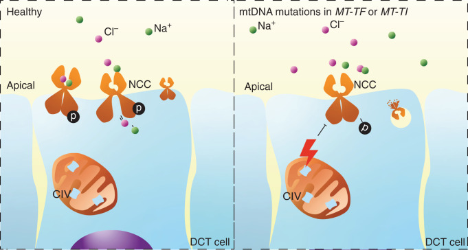
Background: Gitelman syndrome is the most frequent hereditary salt-losing tubulopathy characterized by hypokalemic alkalosis and hypomagnesemia. Gitelman syndrome is caused by biallelic pathogenic variants in SLC12A3, encoding the Na+-Cl- cotransporter (NCC) expressed in the distal convoluted tubule. Pathogenic variants of CLCNKB, HNF1B, FXYD2, or KCNJ10 may result in the same renal phenotype of Gitelman syndrome, as they can lead to reduced NCC activity. For approximately 10 percent of patients with a Gitelman syndrome phenotype, the genotype is unknown.

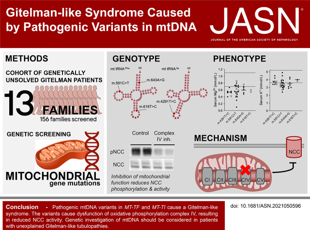
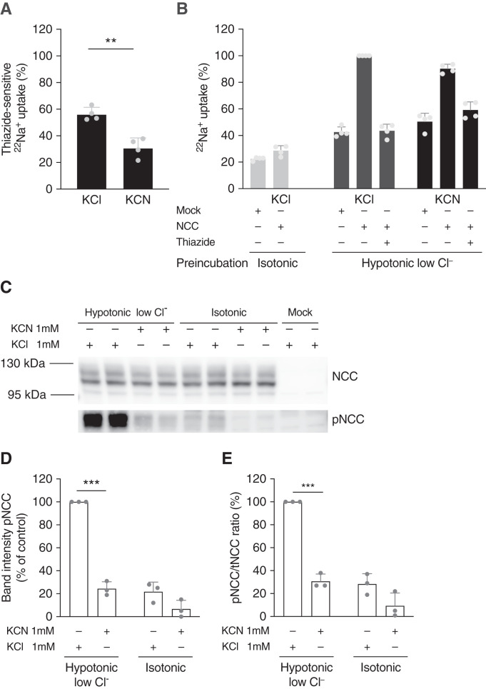
Methods: We identified mitochondrial DNA (mtDNA) variants in three families with Gitelman-like electrolyte abnormalities, then investigated 156 families for variants in MT-TI and MT-TF, which encode the transfer RNAs for phenylalanine and isoleucine. Mitochondrial respiratory chain function was assessed in patient fibroblasts. Mitochondrial dysfunction was induced in NCC-expressing HEK293 cells to assess the effect on thiazide-sensitive 22Na+ transport.

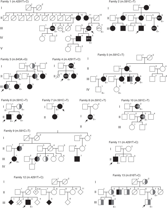
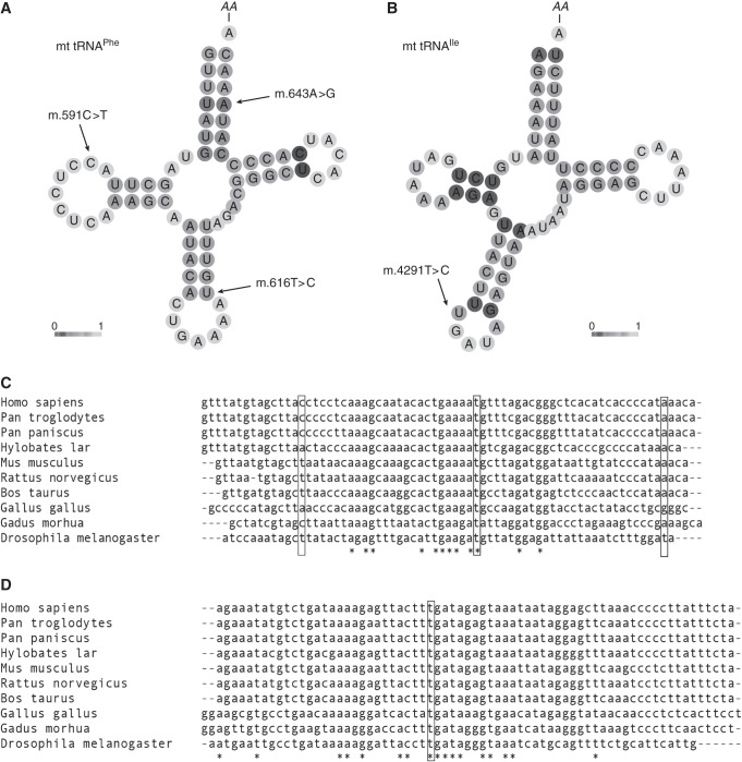
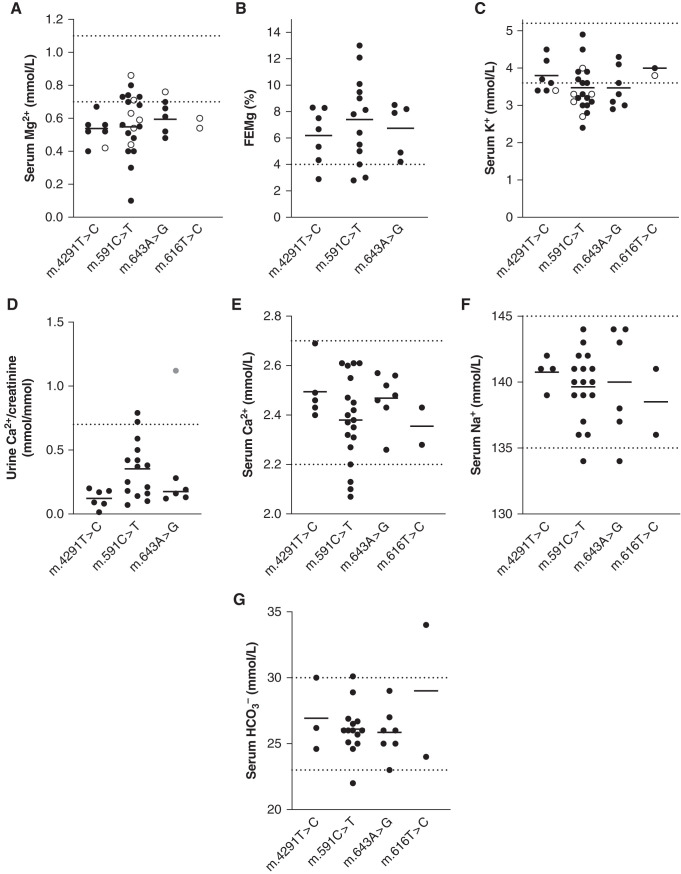
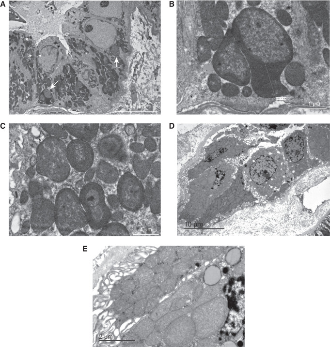
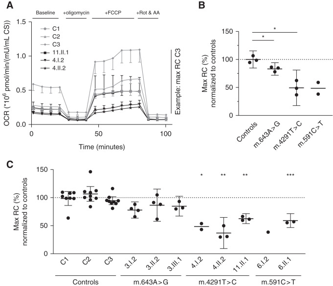
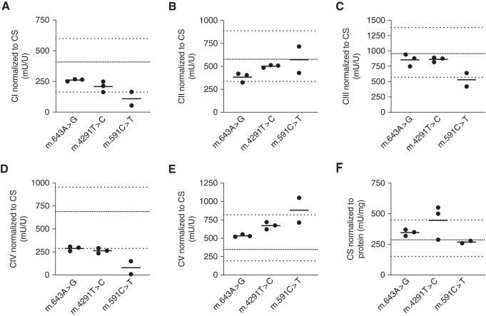
Results: Genetic investigations revealed four mtDNA variants in 13 families: m.591C>T (n=7), m.616T>C (n=1), m.643A>G (n=1) (all in MT-TF), and m.4291T>C (n=4, in MT-TI). Variants were near homoplasmic in affected individuals. All variants were classified as pathogenic, except for m.643A>G, which was classified as a variant of uncertain significance. Importantly, affected members of six families with an MT-TF variant additionally suffered from progressive chronic kidney disease. Dysfunction of oxidative phosphorylation complex IV and reduced maximal mitochondrial respiratory capacity were found in patient fibroblasts. In vitro pharmacological inhibition of complex IV, mimicking the effect of the mtDNA variants, inhibited NCC phosphorylation and NCC-mediated sodium uptake.

Conclusion: Pathogenic mtDNA variants in MT-TF and MT-TI can cause a Gitelman-like syndrome. Genetic investigation of mtDNA should be considered in patients with unexplained Gitelman syndrome-like tubulopathies.

Keywords: Gitelman-s syndrome; Na transport; blood pressure; chronic kidney disease; chronic kidney failure; epithelial sodium transport; genetic renal disease; human genetics; ion transport; mitochondria.


Figures

None
Graphical abstract
Figure 1.
Figure 1.
Pedigrees of the 13 affected families demonstrate maternal inheritance pattern. Black filling denotes tubulopathy. Probands are denoted with arrows; CKD (any stage) is denoted by gray filling. Percentages indicate heteroplasmy level of the variant in blood. E+, the presence of the variant as confirmed by genetic testing; E−, the exclusion of the variant.
Figure 2.
Figure 2.
In silico prediction analysis of variants. In silico prediction analysis of variants in the mt-tRNAs for phenylalanine and isoleucine (mt-tRNAPhe and mt-tRNAIle, respectively). (A and B) CentroidHomFold predictions of secondary structure of the two tRNAs. The grayscale indicates pseudo base-pairing probabilities; light shading represents a low probability and dark shading a high probability. Bold letters indicate anticodons. AA indicates amino acid binding position. (A) Predicted secondary structure of mt-tRNAPhe; the locations of the patient variants m.591C>T, m.616T>C, and m.643A>G are indicated. (B) Predicted secondary structure of mt-tRNAIle; the location of the patient variant m.4291T>C is indicated. (C) MT-TF and (D) MT-TI nucleotide sequences in a standard set of species. Fully conserved residues are indicated by stars (aligned with clustal O).
Figure 3.
Figure 3.
Patients with variants in MT-TI or MT-TF have Gitelman syndrome-like electrolyte abnormalities. (A–G) Serum and urinary electrolyte values in patients by pathogenic variant. Dotted lines represent upper and lower limits of normal. For the fractional excretion of magnesium (FEMg) and urinary calcium excretion, lower limits of normal were not available; therefore, only the upper limit of normal is depicted in panels (B) and (D). Upper limit of normal for FEMg applies to hypomagnesemic individuals only and these are on the basis of Elisaf et al. Black circles (•), without supplementation; open circles (^), with supplementation; gray circle in panel (D), a child (individual 3.III.2); upper limit of normal for this age is 2.2 mmol/mmol Ca2+/creatinine. FEMg is calculated by: serum creatinine×urinary magnesium/(serum magnesium×urinary creatinine)×100%.
Figure 4.
Figure 4.
Renal biopsies of two MT-TF patients show abnormal mitochondria in distal parts of the nephron. (A–C) Transmission electron microscopy of the renal biopsy sample of patient 3.III.1. (D and E) Transmission electron microscopy of a percutaneous renal biopsy sample of patient 10.II.3. (A) Representative image of a perpendicular cross-section of the distal tubule, with a large number of abnormally shaped and sized mitochondria (two examples indicated with white arrows). Cristae profiles appear distorted, including some mitochondria with no discernable cristae. Nanotunneling visible (three examples indicated with white arrowheads). Magnification, ×1000. (B) Close-up of atypical giant mitochondrion of >3 µm in length (same as indicated by the left arrow in panel [A]). Note the large size and compartmentalization. Magnification, ×6000. (C) Close-up of atypical mitochondria (not in panel [A]). Note the concentric cristae (onion-like appearance). Magnification, ×6000. (D) Representative image of a perpendicular cross-section of the distal tubule; enlarged mitochondria are visible. (E) A close-up of panel (D) shows an almost complete lack of cristae structure in most mitochondria.
Figure 5.
Figure 5.
Mitochondrial maximal respiratory capacity is reduced in patient fibroblasts. Mitochondrial function assessed by the Seahorse XFe96 analyzer. (A) Representative OCR plot of a Mito Stress Test of fibroblasts from three patients with the m.4291T>C variant and three controls (n=6 wells for each measurement point). Error bars denote + or − SD. (B) Average maximal mitochondrial respiration for the different mtDNA variants. Each point represents the average of all independent experiments for one individual (n=1–9, depending on the individual, as can be seen in panel [C]). (C) Average maximal mitochondrial respiration for each individual. Each point represents the average of all replicate wells on one Seahorse plate (n=6). (B and C) Means are represented by horizontal bars, error bars denote the 95% confidence interval, and a one-way ANOVA with Dunnett T3 was used to calculate significance. OCR is in pmol O2/min per mU/ml citrate synthase. AA, antimycin A; CS, citrate synthase activity; FCCP, carbonyl cyanide 4‐(trifluoromethoxy)phenylhydrazone; RC, respiratory capacity; Rot, rotenone. *P<0.05; **P<0.005; ***P<0.0005.
Figure 6.
Figure 6.
OXPHOS complex IV activity is reduced in patient mitochondria. (A–E) Activities of the five OXPHOS complexes (CI–CV), and (F) citrate synthase activity. All measurements were performed in isolated mitochondria from patient-derived fibroblasts. Thick, dotted lines represent the reference range from our center; thin, dotted lines represent the means of control individuals. CI to CV, OXPHOS complexes I to V; CS, citrate synthase activity.
Figure 7.
Figure 7.
NCC-mediated sodium uptake and NCC-phosphorylation are reduced with complex IV inhibition. (A and B) 22Na+ uptake in HEK293 cells transfected with NCC or mock, with or without inhibition of OXPHOS complex IV with KCN. KCN 1 mmol/L or KCl 1 mmol/L (control) was added during both preincubation and the uptake period, as indicated; the same applies to hydrochlorothiazide (HCTZ) 100 µmol/L. Bars represent mean with SD. (A) HCTZ-sensitive 22Na+ uptake of NCC-transfected cells over a period of 30 minutes. Data in (A) are on the basis of (B). Significance was assessed with an unpaired t test. (B) 22Na+ uptake in 30 minutes after preincubation with hypotonic-low-chloride buffer or isotonic buffer. Cells were transfected with NCC or mock and treated with KCl or KCN (n=4 of triplicates in each experiment). (C) Representative immunoblots showing NCC and phosphorylated NCC after a 30-minute incubation in hypotonic-low-chloride or isotonic buffer, with either KCN or KCl treatment. The mock condition has been incubated in hypotonic-low-chloride buffer as well. (D–E) Densitometry analysis of pNCC band intensity, and pNCC/tNCC ratio (n=3 duplicates in each experiment). Significance was assessed with unpaired t tests and corrected for multiple testing. pNCC, NCC phosphorylated at Thr60. **P<0.005; ****P<0.00005.
Figure 8.
Figure 8.
Induction of Gitelman-like syndrome by pathogenic mtDNA variants, proposed mechanism. Proposed mechanism of Gitelman-like syndrome induced by pathogenic mtDNA variants in the genes encoding the isoleucine and phenylalanine tRNAs (MT-TI and MT-TF, respectively). The m.4291T>C, m.591C>T, m.616T>C, and m.643A>G variants lead to complex IV dysfunction and reduced maximal respiration. This leads to a decrease in the phosphorylation of NCC and sodium transport. Reduced sodium transport in the DCT leads to reduced magnesium transport in the DCT and increased sodium transport in the collecting duct. Increased sodium reabsorption in the collecting duct leads to increased potassium excretion through ROMK (not shown here). CIV, OXPHOS complex IV.

Comment in

References

    1. Simon DB, Nelson-Williams C, Bia MJ, Ellison D, Karet FE, Molina AM, et al. : Gitelman’s variant of Bartter’s syndrome, inherited hypokalaemic alkalosis, is caused by mutations in the thiazide-sensitive Na-Cl cotransporter. Nat Genet 12: 24–30, 1996 - PubMed
    1. Blanchard A, Bockenhauer D, Bolignano D, Calò LA, Cosyns E, Devuyst O, et al. : Gitelman syndrome: consensus and guidance from a Kidney Disease: Improving Global Outcomes (KDIGO) Controversies Conference. Kidney Int 91: 24–33, 2017 - PubMed
    1. Vargas-Poussou R, Dahan K, Kahila D, Venisse A, Riveira-Munoz E, Debaix H, et al. : Spectrum of mutations in Gitelman syndrome. J Am Soc Nephrol 22: 693–703, 2011 - PMC - PubMed
    1. Downie ML, Lopez Garcia SC, Kleta R, Bockenhauer D: Inherited tubulopathies of the kidney: insights from genetics. Clin J Am Soc Nephrol 16: 620–630, 2020 - PMC - PubMed
    1. Viering DHHM, de Baaij JHF, Walsh SB, Kleta R, Bockenhauer D: Genetic causes of hypomagnesemia, a clinical overview. Pediatr Nephrol 32: 1123–1135, 2017 - PMC - PubMed

Publication types

MeSH terms