Review
. 2021 Oct 4;8(6):e1084.
doi: 10.1212/NXI.0000000000001084. Print 2021 Nov.

Neurosarcoidosis: Pathophysiology, Diagnosis, and Treatment

Affiliations
Review

Neurosarcoidosis: Pathophysiology, Diagnosis, and Treatment

Michael J Bradshaw et al. Neurol Neuroimmunol Neuroinflamm. .

Abstract

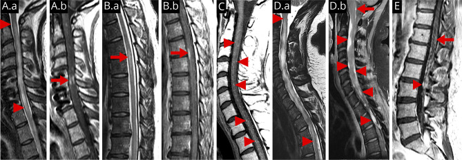
Although often regarded as a protean illness with myriad clinical and imaging manifestations, neurosarcoidosis typically presents as recognizable syndromes that can be approached in a rational, systematic fashion. Understanding of neurosarcoidosis has progressed significantly in recent years, including updated diagnostic criteria and advances in treatment. The diagnosis of neurosarcoidosis is established by the clinical syndrome, imaging and histopathological findings, and exclusion of other causes. Mounting evidence supports the use of tumor necrosis factor inhibitors as an important addition to the therapeutic armamentarium, along with glucocorticoids and steroid-sparing cytotoxic immunosuppressants. In this narrative review, we summarize recent advances in the diagnosis and treatment of neurosarcoidosis.


Figures

Figure 1
Figure 1. Brain MRI Manifestations of Neurosarcoidosis
Typical patterns of NS observed on brain MRI including leptomeningeal and pachymeningeal disease, perivascular infiltration, MS-like lesions, which may represent coexistent disease, mass-like lesions, cranial nerve infiltration, and pituitary/meningeal disease. (A.a–A.b) Post-gadolinium T1 MRI from a patient with probable NS demonstrating leptomeningeal enhancement predominantly affecting the base of the brain (arrows). (B.a) T2/FLAIR MRI of definite NS demonstrates T2 hyperintensity in the occipital lobe (arrow) adjacent to an area of (B.b) dural thickening and enhancement (arrow) on post-gadolinium T1 MRI. (C) Post-gadolinium T1 MRI demonstrating multifocal perivascular meningeal enhancement in the subcortical white matter (arrows) and cortex (arrowhead) in definite NS. (D) Sagittal FLAIR MRI of the same patient from B, exhibited T2 hyperintense lesions in the cerebellum (not shown) as well as the periventricular and juxtacortical white matter (arrows). Atrophy associated with the occipital lobe T2 hyperintense lesion can be appreciated (arrowhead). This patient was diagnosed with multiple sclerosis and later developed pachymeningeal thickening biopsy proven to be sarcoidosis. (E.a) Post-gadolinium T1 MRI demonstrating a focal area of contrast enhancement in the left insula (arrow) with (E.b) parenchymal T2 hyperintensity on FLAIR in a patient with probable neurosarcoidosis. (F.a) Mass-like lesion in the right mesial temporal lobe with enhancement (arrow) on post-gadolinium T1 MRI with corresponding (F.b) swelling and T2 hyperintensity on FLAIR (arrow) in definite NS. Note that most mass-like lesions in NS have some meningeal component as seen in E and F. (G) Coronal post-gadolinium T1 MRI demonstrating left optic nerve enhancement (arrow) in a patient with probable NS who presented with left optic neuritis. (H) Axial post-gadolinium T1 MRI with contrast enhancement and swelling in Meckel cave about the left trigeminal nerve (arrow) in a patient with probable NS who developed left trigeminal neuropathy (pain and decreased sensation). (I) Axial post-gadolinium T1 MRI demonstrating contrast enhancement in both internal auditory meatuses (arrows) in a patient with probable NS and bilateral facial nerve palsy. (J) Sagittal post-gadolinium T1 MRI in a patient with definite NS who presented with hypopituitarism demonstrates abnormal contrast enhancement and swelling of the pituitary gland (arrowhead) and stalk (arrow).
Figure 2
Figure 2. Spinal Cord MRI Manifestations of Neurosarcoidosis
Typical patterns of spinal cord involvement in NS including longitudinally extensive myelitis, short segment myelitis, perivascular and leptomeningeal lesions, mas-like dural and leptomeningeal lesions, and lumbosacral radiculitis. (A.a) Sagittal T2 MRI of the cervical spine demonstrating longitudinally extensive transverse myelitis (between arrowheads) in a patient with probable NS and (A.b) extensive contrast enhancement on post-gadolinium T1 MRI (arrow). (B.a) Sagittal T2 MRI of the thoracic spine demonstrating a short segment myelitis (arrow) in a patient with probable NS with (B.b) contrast enhancement on post-gadolinium T1 MRI (arrow). (C) Sagittal post-gadolinium T1 MRI of the cervical spine from the same patient in Figure 1C demonstrating multifocal areas of contrast enhancement (arrowheads) in an unusual pattern suggesting leptomeningeal and perivascular infiltration. (D.a) Sagittal T2 MRI of the cervical spine with longitudinally extensive T2 hyperintensity and cord deformity in a patient with definite NS. (D.b) Sagittal post-gadolinium T1 MRI from the same patient in (D.a) demonstrating multiple mass-like enhancing lesions surrounding and compressing the spinal cord (arrowheads) well as leptomeningeal enhancement about the medulla (arrow). (E) Sagittal post-gadolinium T1 MRI of the lumbar spine demonstrating nodular leptomeningeal enhancement about the cauda equina (arrowhead) and enhancement within the conus medullaris (arrow) in a patient with definite NS who presented with lumbosacral radiculomyelitis (Elsberg syndrome).

References

    1. Valeyre D, Prasse A, Nunes H, Uzunhan Y, Brillet PY, Müller-Quernheim J. Sarcoidosis. Lancet. 2014;383(9923):1155-1167. - PubMed
    1. Rossi G, Cavazza A, Colby TV. Pathology of sarcoidosis. Clin Rev Allergy Immunol. 2015;49(1):36-44. - PubMed
    1. Ungprasert P, Carmona EM, Utz JP, Ryu JH, Crowson CS, Matteson EL. Epidemiology of sarcoidosis 1946-2013: a population-based study. Mayo Clin Proc. 2016;91(2):183-188. - PMC - PubMed
    1. Joubert B, Chapelon-Abric C, Biard L, et al. . Association of prognostic factors and immunosuppressive treatment with long-term outcomes in neurosarcoidosis. JAMA Neurol. 2017;74(11):1336-1344. - PMC - PubMed
    1. Fritz D, van de Beek D, Brouwer MC. Clinical features, treatment and outcome in neurosarcoidosis: systematic review and meta-analysis. BMC Neurol. 2016;16(1):220. - PMC - PubMed

MeSH terms

Supplementary concepts