. 2021 Oct 12;118(41):e2107771118.
doi: 10.1073/pnas.2107771118.

Eicosanoid regulation of debris-stimulated metastasis

Affiliations

Eicosanoid regulation of debris-stimulated metastasis

Jianjun Deng et al. Proc Natl Acad Sci U S A. .

Abstract

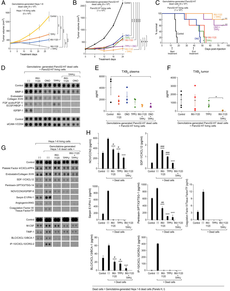
Cancer therapy reduces tumor burden via tumor cell death ("debris"), which can accelerate tumor progression via the failure of inflammation resolution. Thus, there is an urgent need to develop treatment modalities that stimulate the clearance or resolution of inflammation-associated debris. Here, we demonstrate that chemotherapy-generated debris stimulates metastasis by up-regulating soluble epoxide hydrolase (sEH) and the prostaglandin E2 receptor 4 (EP4). Therapy-induced tumor cell debris triggers a storm of proinflammatory and proangiogenic eicosanoid-driven cytokines. Thus, targeting a single eicosanoid or cytokine is unlikely to prevent chemotherapy-induced metastasis. Pharmacological abrogation of both sEH and EP4 eicosanoid pathways prevents hepato-pancreatic tumor growth and liver metastasis by promoting macrophage phagocytosis of debris and counterregulating a protumorigenic eicosanoid and cytokine storm. Therefore, stimulating the clearance of tumor cell debris via combined sEH and EP4 inhibition is an approach to prevent debris-stimulated metastasis and tumor growth.

Keywords: autacoid; debris; inflammation resolution; prostaglandin E2 receptor 4; soluble epoxide hydrolase.


Conflict of interest statement

Competing interest statement: Y.S., K.-K.H., X.Y., and B.D.H. work with Ionova and EicOsis, respectively, on the development of EP4 and epoxide hydrolase inhibitors for clinical use. D.P., A.G., and B.D.H., and colleagues C. N. Serhan and P. Sime were coauthors on an earlier commentary suggesting modulating the eicosanoid and cytokine storms with a soluble epoxide hydrolase inhibitor to modulate severity of COVID infections, introducing the same mechanism as is suggested here for reduction of the cytokine storm following cancer therapy.

Figures

Fig. 1.
Fig. 1.
Chemotherapy-generated tumor cell debris stimulates pancreatic cancer via upregulation of sEH and EP4. (A) Pancreatic tumor growth stimulated by gemcitabine-generated Panc02-H7 debris (9 × 10⁵ dead cells) coinjected with a subthreshold inoculum of Panc02-H7 (1 × 10⁴ living cells). n = 5 mice per group. ***P < 0.001 vs. dead cells or living cells (“no dead cells”) alone. Relative gene expression of (B) Ephx2 (sEH) and (C) Ptger4 (EP4) in debris-stimulated tumor tissue (9 × 10⁵ gemcitabine-generated Panc02-H7 dead cells + 1 × 10⁴ Panc02-H7 living cells) vs. control tumor (1 × 10⁴ Panc-02-H7 living cells). mRNA expression levels of genes were analyzed by qRT-PCR and normalized by GAPDH. n = 3 per group. *P < 0.05, **P < 0.01 vs. control.
Fig. 2.
Fig. 2.
The cytokine storm triggered by debris-stimulated macrophages is prevented by combined sEH and EP4 inhibition. (A and B) Angiogenic (Upper) and inflammatory (Lower) cytokines from conditioned medium of RAW 264.7 macrophages treated with vehicle, sEH inhibitor (EC5026 or TPPU, 10 µM, 2 h), EP4 antagonist (INV-1120 or ONO-AE3-208, 10 µM, 2 h), or a combination (EC5026 + ONO-AE3-208, TPPU + ONO-AE3-208, or TPPU + INV-1120, 10 µM each, 2 h) and subsequently stimulated by gemcitabine-generated Panc02-H7 tumor cell debris vs. no debris. The SDF-1/CXCL12 released from RAW 264.7 macrophages was quantified by ELISA. Data are presented as means (pg/mL) ± SEM n = 3 per group. *P < 0.05, **P < 0.01, ***P < 0.001 vs. dead cells + Panc02-H7 living cells. #P < 0.05, ###P < 0.001 vs. TPPU + ONO-AE3-208. (C and D) Angiogenic (Upper) and inflammatory (Lower) cytokines from conditioned medium of RAW 264.7 macrophages treated with vehicle, sEH inhibitor (EC5026 or TPPU, 10 µM, 2 h), EP4 antagonist (INV-1120 or ONO-AE3-208, 10 µM, 2 h), or a combination (EC5026 + ONO-AE3-208, TPPU + ONO-AE3-208, or TPPU + INV-1120, 10 µM each, 2 h) and subsequently stimulated by gemcitabine-generated Hepa 1-6 tumor cell debris vs. no debris. The IGFBP-1 and VEGF/VPF released from RAW 264.7 macrophages were quantified by ELISA. Data are presented as means (pg/mL) ± SEM n = 3 per group. **P < 0.01, ***P < 0.001 vs. dead cells + Hepa 1-6 living cells. #P < 0.05, ##P < 0.01, ###P < 0.001. (E and F) Angiogenic (Upper) and inflammatory (Lower) cytokines from conditioned medium of hMDMs treated with vehicle, sEH inhibitor (EC5026 or TPPU, 10 µM, 2 h), EP4 antagonist (INV-1120, 10 µM, 2 h), or a combination (EC5026 + INV-1120 or TPPU + INV-1120, 10 µM each, 2 h) and subsequently stimulated by gemcitabine-generated HepG2 tumor cell debris vs. no debris.
Fig. 3.
Fig. 3.
Combined inhibition of sEH and EP4 stimulates macrophage phagocytosis of debris via suppression of NF-κB signaling. Combination treatment with an sEH inhibitor (EC5026 or TPPU, 10 µM) and an EP4 antagonist (INV-1120 or ONO-AE3-208, 10 µM) for 2 h stimulates (A) RAW 264.7 murine macrophage phagocytosis or (B) hMDM phagocytosis of CFDA-labeled gemcitabine-generated Hepa 1-6 or HepG2 tumor cell debris. Macrophage phagocytosis was measured as RFU and normalized to percent increase above vehicle-treated macrophages. n = 12 per group. *P < 0.05, ***P < 0.001; #P < 0.05, ##P < 0.01, ###P < 0.001 vs. vehicle. (C) Western blot analysis of p-IKKβ, IKKβ, p-NF-κB, NF-κB, IκBα, p-AKT, and AKT in living or debris-stimulated Hepa 1-6 tumors from mice treated for 28 d with vehicle, INV-1120 (5 mg/kg/d), TPPU (5 mg/kg/d), or INV-1120 + TPPU (5 mg/kg/d each). β-Actin was used as a loading control. (D) Quantification of protein expression shown in C. n = 3 mice per group. *P < 0.05.
Fig. 4.
Fig. 4.
Combined inhibition of sEH and EP4 differentially regulates the release of eicosanoids by debris-stimulated macrophages. LC-MS/MS–based oxylipin analysis of conditioned medium from RAW 264.7 macrophages stimulated by gemcitabine-generated (A) Panc02-H7 or (B) Hepa 1-6 debris, macrophages treated with an sEH inhibitor (TPPU, 10 µM, 2 h) and/or EP4 antagonist (INV-1120, 10 µM, 2 h) and stimulated by gemcitabine-generated Panc02-H7 debris, gemcitabine-generated Hepa 1-6 debris, or macrophages not stimulated with debris (“macrophages [MØ] alone”). (A and B) Heat maps; (CJ) Quantification of 8 oxylipins in A and B, including PGE2, 15-deoxy-PGJ2, 11-HETE, 15(S)-HETE, 9-HETE, 8 (9)-EpETrE, 11,12-DiHETrE, and 5,6-DiETrE. Data are presented as means (pg/mL) ± SEM n = 3 per group. *P < 0.05, **P < 0.01, ***P < 0.001, n.s., not significant.
Fig. 5.
Fig. 5.
Prevention of debris-stimulated liver metastasis and cytokine storm by sEH inhibition and EP4 antagonism. (A) Growth of debris-stimulated tumors [gemcitabine-generated Hepa 1-6 debris [9 × 10⁵ dead cells] + Hepa 1-6 [5 × 10⁶ living cells]) systemically treated with TPPU (5 mg/kg/d), INV-1120 (5 mg/kg/d), or TPPU + INV-1120 (5 mg/kg/d each). Treatment initiated once tumors reached 100 to 200 mm3. n = 5 mice per group. **P < 0.01, ***P < 0.001. (B) Growth of debris-stimulated tumors (gemcitabine-generated Panc02-H7 debris [9 × 10⁵ dead cells] + Panc02-H7 [1 × 10⁴ living cells]) systemically treated with an sEH inhibitor (TPPU, 5 mg/kg/d), EP4 antagonist (INV-1120 or ONO-AE3-208, 5 mg/kg/d), or a combination of them (TPPU + INV-1120 or TPPU + ONO-AE3-208, 5 mg/kg/d each). n = 5 mice per group. *P < 0.05, **P < 0.01, ***P < 0.001. (C) Percent survival of mice coinjected orthotopically with gemcitabine-generated Panc02-H7 debris (9 × 10⁵ dead cells) and Panc02-H7 (1 × 10⁴ living cells). Mice were systemically treated with an sEH inhibitor (TPPU, 5 mg/kg/d), EP4 antagonist (INV-1120 or ONO-AE3-208, 5 mg/kg/d), or a combination (TPPU + INV-1120, TPPU + ONO-AE3-208, or TPPU + INV-1120, 5 mg/kg/d each). Kaplan–Meier analysis indicated significantly prolonged survival in treated mice compared to control. n = 5 mice per group. *P < 0.05, **P < 0.01 vs. control. (D) Angiogenic (Upper) and inflammatory (Lower) cytokines in plasma, as well as TXB2 in plasma (E) or tumor tissues (F), from mice bearing debris-stimulated subcutaneous Panc02-H7 tumors treated with TPPU, INV-1120, ONO-AE3-208, TPPU + INV-1120, or TPPU + ONO-AE3-208. Plasma and tumor tissues were collected on treatment day 24. TXB2 product was quantified by ELISA. Data are presented as means (pg/mL) ± SEM n = 3 per group. *P < 0.05. (G) Angiogenic (Upper) and inflammatory (Lower) cytokines of plasma from mice bearing debris-stimulated subcutaneous Hepa 1-6 tumors treated with TPPU, INV-1120, or TPPU + INV-1120. Plasma was collected on treatment day 28. (H) Quantification of cytokines by ELISA shown in G. Data were presented as means (pg/mL) ± SEM n = 3 per group. *P < 0.05, **P < 0.01, ***P < 0.001 vs. debris-stimulated tumors without treatment. #P < 0.05, ##P < 0.01, ###P < 0.001 vs. INV-1120 + TPPU.
Fig. 6.
Fig. 6.
Potential mechanism for the resolution of debris-stimulated metastatic hepato-pancreatic cancer via combined soluble epoxide hydrolase and EP4 inhibition.

Similar articles

Cited by

References

    1. Pinheiro P. S., et al. ., Liver cancer: A leading cause of cancer death in the United States and the role of the 1945-1965 birth cohort by ethnicity. JHEP Rep 1, 162–169 (2019). - PMC - PubMed
    1. Von Hoff D. D., et al. ., Increased survival in pancreatic cancer with nab-paclitaxel plus gemcitabine. N. Engl. J. Med. 369, 1691–1703 (2013). - PMC - PubMed
    1. Abubaker K., et al. ., Short-term single treatment of chemotherapy results in the enrichment of ovarian cancer stem cell-like cells leading to an increased tumor burden. Mol. Cancer 12, 24 (2013). - PMC - PubMed
    1. Gunjal P. M., et al. ., Evidence for induction of a tumor metastasis-receptive microenvironment for ovarian cancer cells in bone marrow and other organs as an unwanted and underestimated side effect of chemotherapy/radiotherapy. J. Ovarian Res. 8, 20 (2015). - PMC - PubMed
    1. Volk-Draper L., et al. ., Paclitaxel therapy promotes breast cancer metastasis in a TLR4-dependent manner. Cancer Res. 74, 5421–5434 (2014). - PMC - PubMed

Publication types

MeSH terms