Review
. 2021 Oct 4;134(22):2647-2655.
doi: 10.1097/CM9.0000000000001772.

Molecular mechanism and therapeutic targeting of necrosis, apoptosis, pyroptosis, and autophagy in cardiovascular disease

Affiliations
Review

Molecular mechanism and therapeutic targeting of necrosis, apoptosis, pyroptosis, and autophagy in cardiovascular disease

Pan Li et al. Chin Med J (Engl). .

Abstract

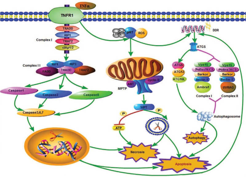
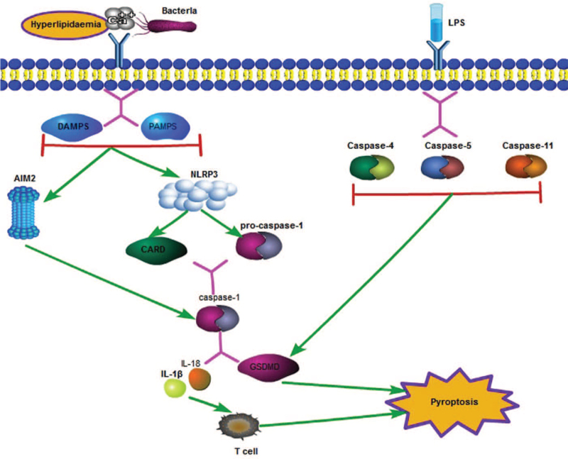
Cell death occurs in various tissues and organs in the body. It is a physiological or pathological process that has different effects. It is of great significance in maintaining the morphological function of cells and clearing abnormal cells. Pyroptosis, apoptosis, and necrosis are all modes of cell death that have been studied extensively by many experts and scholars, including studies on their effects on the liver, kidney, the heart, other organs, and even the whole body. The heart, as the most important organ of the body, should be a particular focus. This review summarizes the mechanisms underlying the various cell death modes and the relationship between the various mechanisms and heart diseases. The current research status for heart therapy is discussed from the perspective of pathogenesis.


Conflict of interest statement

None.

Figures

Figure 1
Figure 1
The mechanisms underlying apoptosis, necrosis, and autophagy. In the mitochondrial pathway of necrosis, intracellular and extracellular Ca2+, alkaline pH, and ROS can stimulate the opening of the MPTP (composed of the voltage-dependent anion channel, adenine nuclear translocator [ANT], and cyclin D). Oxidative phosphorylation leads to the cessation of ATP production and destruction of the cell membrane, leading to necrosis. This process can also lead to apoptosis. When the death receptor pathway is triggered, the binding of TNF to TNFR1 leads to exposure of the cytoplasmic death domain of TNFR1, which in turn leads to the recruitment of TRADD. It provides a scaffold for the assembly of complex I (TNFR1, TRADD, RIP1, TRAF2, and cIAP1/2) on the plasma membrane. The complex can promote the formation of complex II (FADD, TRADD, RIP1/3, and TRAF2) under certain conditions. Complex II can mediate the recruitment and activation of caspase-1/2/8, and then activate the downstream caspase-3/6/7. These caspase proteins can destroy DNA in the nucleus, and lead to the destruction of a large number of plasma and cell membranes, thereby leading to necrosis. The activation of caspase proteins, especially caspase-3, is the key factor for triggering apoptosis. DDR induced by various factors leads to the formation of ATGs, such as ATG5 and Beclin-1. ATG5 mediates the formation of the autophagosome through the formation of ATG5–ATG1–ATG16L, while Beclin-1 mediates the formation of the autophagosome through the formation of complex I (composed of Beclin-1, PtdIns3KC3, Vps15, Bakor, and Ambra1) and complex II (composed of Beclin-1, PtdIns3KC3, Vps15, Bakor, and UVRAG), which mediate the formation of autophagosomes, eventually leading to autophagy. The process of apoptosis also depends on the formation of ATGs. ATGs: Autophagy-related proteins; cIAP1/2: Cellular inhibitors of apoptosis 1/2; DDR: DNA damage response; FADD: Fas-associated via death domain; MPTP: Mitochondrial permeability transition pore; PtdIns3KC3: Phosphatidylinositol 3-kinase; ROS: Reactive oxygen species; TNF: Tumor necrosis factor; TNFR1: TNF receptor1; TRADD: TNF-receptor-associated death domain; TRAF2: TNF-related apoptosis-inducing ligand receptors.
Figure 2
Figure 2
Caspase-1-dependent and independent signaling pathways of pyroptosis. In the classical pathway, when the body is stimulated by hyperlipidemia, abnormal Ca2+ levels, and bacteria, infection cells activate downstream inflammatory bodies, including NLRP3 and AIM2 through PAMP and DAMP pathways. NLRP3 can establish a link with CARD and procaspase-1. Procaspase-1 bound to CARD can self-cleave into mature caspase-1, and AIM2 can directly activate caspase-1. Mature caspase-1 can cleave the downstream GSDMD and can induce large production of IL-18 and other inflammatory factors. The N-terminal of GSDMD, which is cleaved, will combine with phospholipids and protein on the cell membrane to form pores, leading to the outflow of inflammatory factors and cell contents. Inflammatory factors can activate T cell immunity in the body, further aggravate body damage, and eventually lead to cell death. The non-classical pathway of pyroptosis can be induced by LPS, which can activate caspase-4/5/11, and also lead to GSDMD cleavage. AIM2: Absent in melanoma 2; CARD: Caspase activation and recruitment domain; DAMP: Danger-associated molecular patterns; GSDMD: Gasdermin D; IL: Interleukin; LPS: Lipopolysaccharides; NLRP3: Nucleotide-binding oligomerization domain-like receptor protein 3; PAMP: Pathogen-associated molecular pattern.

References

    1. Yamaguchi O, Higuchi Y, Hirotani S, Kashiwase K, Nakayama H, Hikoso S, et al. . Targeted deletion of apoptosis signal-regulating kinase 1 attenuates left ventricular remodeling. Proc Natl Acad Sci U S A 2003; 100:15883–15888. doi: 10.1073/pnas.2136717100. - PMC - PubMed
    1. Rizzuto R, Pozzan T. Microdomains of intracellular Ca2+: molecular determinants and functional consequences. Physiol Rev 2006; 86:369–408. doi: 10.1152/physrev.00004.2005. - PubMed
    1. Benjamin EJ, Blaha MJ, Chiuve SE, Cushman M, Das SR, Deo R, et al. . American Heart Association Statistics Committee and Stroke Statistics Subcommittee. Heart disease and stroke statistics-2017 update: a report from the American Heart Association. Circulation 2017; 135:e146–e603. doi: 10.1161/CIR.0000000000000485. - PMC - PubMed
    1. Nag AC. Study of non-muscle cells of the adult mammalian heart: a fine structural analysis and distribution. Cytobios 1980; 28:41–61. - PubMed
    1. Bulluck H, Rosmini S, Abdel-Gadir A, White SK, Bhuva AN, Treibel TA, et al. . Residual myocardial iron following intramyocardial hemorrhage during the convalescent phase of reperfused ST-segment-elevation myocardial infarction and adverse left ventricular remodeling. Circ Cardiovasc Imaging 2016; 9:e004940.doi: 10.1161/CIRCIMAGING.116.004940. - PMC - PubMed