. 2021 Oct 4;11(1):19635.
doi: 10.1038/s41598-021-99086-2.

Leu-to-Phe substitution at prM146 decreases the growth ability of Zika virus and partially reduces its pathogenicity in mice

Affiliations

Leu-to-Phe substitution at prM146 decreases the growth ability of Zika virus and partially reduces its pathogenicity in mice

Takuya Inagaki et al. Sci Rep. .

Abstract

Zika virus (ZIKV) is a mosquito-borne flavivirus that causes febrile illness. The recent spread of ZIKV from Asia to the Americas via the Pacific region has revealed unprecedented features of ZIKV, including transplacental congenital infection causing microcephaly. Amino acid changes have been hypothesized to underlie the spread and novel features of American ZIKV strains; however, the relationship between genetic changes and the epidemic remains controversial. A comparison of the characteristics of a Southeast Asian strain (NIID123) and an American strain (PRVABC59) revealed that the latter had a higher replication ability in cultured cells and higher virulence in mice. In this study, we aimed to identify the genetic region of ZIKV responsible for these different characteristics using reverse genetics. A chimeric NIID123 strain in which the E protein was replaced with that of PRVABC59 showed a lower growth ability than the recombinant wild-type strain. Adaptation of the chimeric NIID123 to Vero cells induced a Phe-to-Leu amino acid substitution at position 146 of the prM protein; PRVABC59 also has Leu at this position. Leu at this position was found to be responsible for the viral replication ability and partially, for the pathogenicity in mouse testes.


Conflict of interest statement

The authors declare no competing interests.

Figures
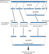
Figure 1
Figure 1
Schematic diagram of the reverse genetics system used for the ZIKV PRVABC59 and ZIKV/Hu/NIID123/2016 (NIID123) strains. For cloning, the whole-genome cDNA of ZIKV was divided into six regions (A1, A2, B, C, D, and E). pMW119 and pMW119-CMVP, which has CMV promoter and HDV ribozyme sequences in the multicloning site of pMW119, were used,. Fragments A1 and A2 were cloned into pMW119-CMVP and pMW119, respectively. Fragments B, C, D, and E were concatenated by joint PCR and then cloned into pMW119-CMVP. Six plasmid clones, pZV-PRVABC59-A1, pZV-PRVABC59-A2, pZV-PRVABC59-BE, pZV-NIID123-A1, pZV-NIID123-A2, and pZV-NIID123-BE, were obtained. To construct the full-length virus cDNA, the fragments in the clones were PCR-amplified and concatenated as full-length viral cDNA by joint PCR. The full-length viral cDNA with the CMV promoter sequence in the 5′-terminus and the HDV ribozyme sequence in the 3′-terminus was transfected into Vero cells to produce infectious viruses.
Figure 2
Figure 2
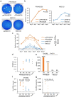
Characterization of recombinant ZIKV rPRVABC59 and rNIID123 in vitro and in vivo. (a) Plaque morphology of the parental virus PRVABC59 (paPRVABC59) and rPRVABC59 (left), and the parental virus NIID123 (paNIID123) and rNIID123 (right) in Vero cells at 5 days post infection. (b) Growth curves of parental and recombinant PRVABC59 (left), and parental and recombinant NIID123 (right) in Vero cells. Cells were inoculated with the viruses at an MOI of 0.01 pfu/cell. Means ± standard deviations of three independent experiments are shown. (c) Viremia levels in mice inoculated with parental and recombinant PRVABC59, and parental and recombinant NIID123. IFNAR1-KO mice (4 or 5 mice/virus group) inoculated with parental or recombinant virus via the footpad were divided into two subgroups (2 or 3 mice/subgroup; 1-, 3-, and 6-days post-infection group and 2-, 4-, and 8-day post-infection group) and blood was collected at the indicated time points. Means + standard deviations are shown. (d) Testis weights at 6 weeks post infection. (e) Ratios of atrophied to total testes in mice infected with the indicated viruses. (f,g) Viral RNA copy numbers in testes (f) and serum (g) at 6 weeks post infection. Dotted line: limit of detection. pa: parental virus. rec: recombinant virus.
Figure 3
Figure 3
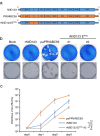
Generation and characterization of chimera ZIKV rNIID123 EPRV in vitro. (a) Schematic representation of the genome structure of rNIID123 EPRV. (b) Plaques (upper panels) and focus phenotypes (lower panels) of rNIID123, paPRVABC59, and rNIID123 EPRV #1 and #2 in Vero cells. (c) Growth curves for the parental virus paPRVABC59, rNIID123, and rNIID123 EPRV #2. Cells were inoculated with the viruses at an MOI of 0.01 ffu/cell. Means ± standard deviations of three independent experiments are shown.
Figure 4
Figure 4
Recovery of Vero cell-adapted rNIID123 EPRV and in vitro growth of the point mutant rNIID123 prMF146L. (a) Focus phenotypes of rNIID123 EPRV #1 after one (P1) to four (P4) passages in Vero cells. (b) Partial amino acid sequences of the prM region in rNIID123 EPRV #1 P1, P4, and PRVABC59. Numbers indicate positions in the ZIKV prM protein. (c) Schematic representation of the point mutant rNIID123 prMF146L. (d) Focus phenotypes of rNIID123 and rNIID123 prMF146L in Vero cells. (e) Growth curves of rNIID123 EPRV P4 and rNIID123 prMF146L in Vero cells. Cells were inoculated with the viruses at an MOI of 0.01 ffu/cell. Means ± standard deviations of three independent experiments are shown.
Figure 5
Figure 5
In vitro growth of the point mutant rPRVABC59 prML146F. (a) Schematic representation of the mutant rPRVABC59 prML146F. (b) Focus phenotypes of rPRVABC59 and rPRVABC59 prML146F in Vero cells. (c) Growth curves of the point mutants rPRVABC59 prML146F and rNIID123 prMF146L in Vero cells. Cells were inoculated with the viruses at an MOI of 0.01 ffu/cell. Means ± standard deviations of three independent experiments are shown.
Figure 6
Figure 6
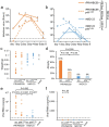
Characterization of the point mutants rPRVABC59 prML146F and rNIID123 prMF146L in mice. (a,b) IFNAR1-KO mice (6 mice/virus group) inoculated with rPRVABC59,rPRVABC59 prML146F, rNIID123 and rNIID123 prMF146L via the footpad were divided into two subgroups (3 mice/subgroup; 1-, 3-, and 6-days post-infection group and 2-, 4-, and 8-days post-infection group) and blood was collected at the indicated time points. Means + standard deviations are shown. (a) rPRVABC59 and rPRVABC59 prML146F. (b) rNIID123 and rNIID123 prMF146L. (c) Testis weight at 6 weeks post infection. (d) Ratios of atrophied to total testes in mice infected with the indicated viruses. (e,f) Viral RNA copy numbers in testes (e) and serum (f) at 6 weeks post infection. Dotted line: limit of detection.
Figure 7
Figure 7
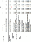
Alignment of partial amino acid sequences of the prM protein of ZIKV strains. Twenty-eight strains representing the African lineage and the Southeast Asian, Pacific, and American subtypes of the Asian/American lineage of ZIKV were used. The amino acid residue at position 146 of the prM protein in the NIID123 strain is indicated by a red rectangle.

Similar articles

Cited by

References

    1. Simmonds P, et al. ICTV virus taxonomy profile: Flaviviridae. J. Gen. Virol. 2017;98:2–3. doi: 10.1099/jgv.0.000672. - DOI - PMC - PubMed
    1. Weaver SC, Reisen WK. Present and future arboviral threats. Antiviral Res. 2010;85:328–345. doi: 10.1016/j.antiviral.2009.10.008. - DOI - PMC - PubMed
    1. Musso D, Ko AI, Baud D. Zika virus infection: After the pandemic. N. Engl. J. Med. 2019;381:1444–1457. doi: 10.1056/NEJMra1808246. - DOI - PubMed
    1. Song B-H, Yun S-I, Woolley M, Lee Y-M. Zika virus: History, epidemiology, transmission, and clinical presentation. J. Neuroimmunol. 2017;308:50–64. doi: 10.1016/j.jneuroim.2017.03.001. - DOI - PubMed
    1. Duffy MR, et al. Zika virus outbreak on Yap Island, federated states of Micronesia. N. Engl. J. Med. 2009;360:2536–2543. doi: 10.1056/NEJMoa0805715. - DOI - PubMed

Publication types

Substances