. 2021 Nov;40(46):6430-6442.
doi: 10.1038/s41388-021-02030-x. Epub 2021 Oct 4.

RNA binding protein RBMS3 is a common EMT effector that modulates triple-negative breast cancer progression via stabilizing PRRX1 mRNA

Affiliations

RNA binding protein RBMS3 is a common EMT effector that modulates triple-negative breast cancer progression via stabilizing PRRX1 mRNA

C James Block et al. Oncogene. 2021 Nov.

Abstract

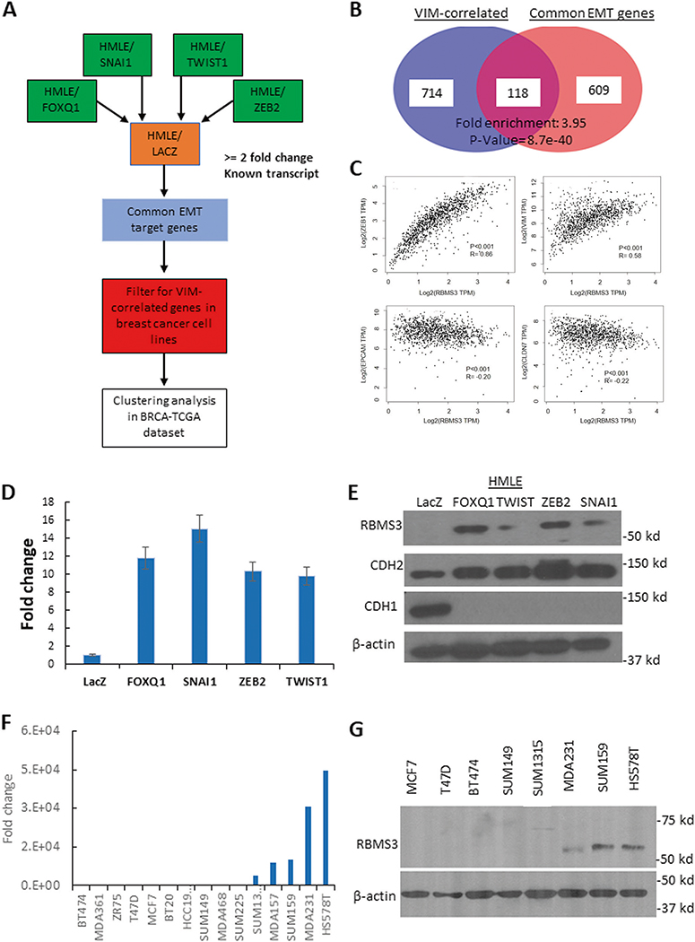
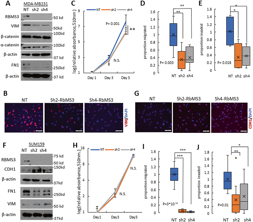
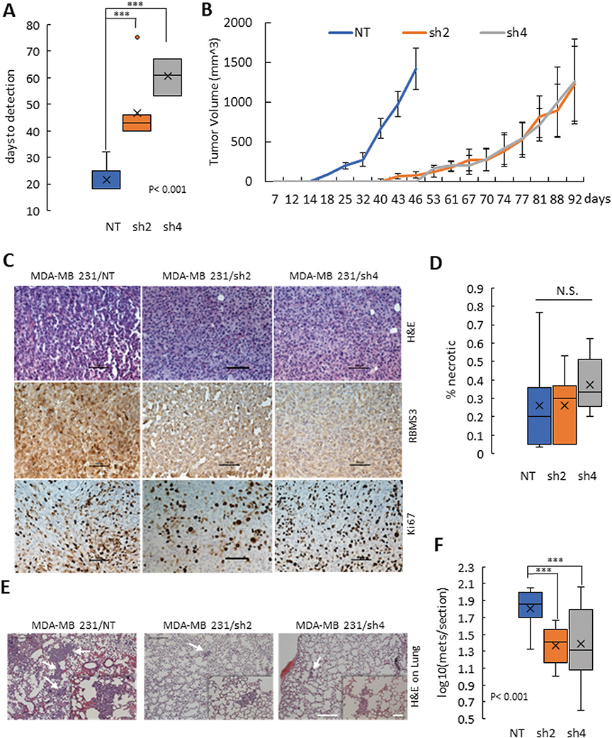
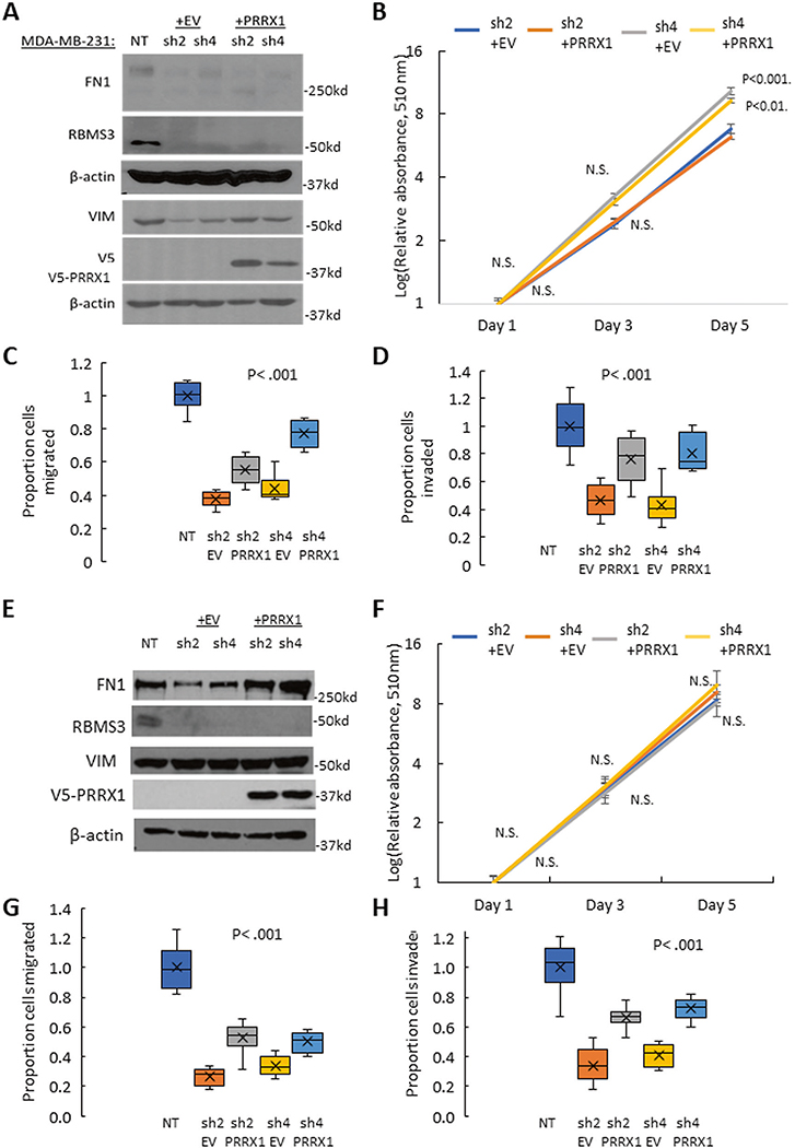
The epithelial-to-mesenchymal transition (EMT) has been recognized as a driving force for tumor progression in breast cancer. Recently, our group identified the RNA Binding Motif Single Stranded Interacting Protein 3 (RBMS3) to be significantly associated with an EMT transcriptional program in breast cancer. Additional expression profiling demonstrated that RBMS3 was consistently upregulated by multiple EMT transcription factors and correlated with mesenchymal gene expression in breast cancer cell lines. Functionally, RBMS3 was sufficient to induce EMT in two immortalized mammary epithelial cell lines. In triple-negative breast cancer (TNBC) models, RBMS3 was necessary for maintaining the mesenchymal phenotype and invasion and migration in vitro. Loss of RBMS3 significantly impaired both tumor progression and spontaneous metastasis in vivo. Using a genome-wide approach to interrogate mRNA stability, we found that ectopic expression of RBMS3 upregulates many genes that are resistant to degradation following transcriptional blockade by actinomycin D (ACTD). Specifically, RBMS3 was shown to interact with the mRNA of EMT transcription factor PRRX1 and promote PRRX1 mRNA stability. PRRX1 is required for RBMS3-mediated EMT and is partially sufficient to rescue the effect of RBMS3 knockdown in TNBC cell lines. Together, this study identifies RBMS3 as a novel and common effector of EMT, which could be a promising therapeutic target for TNBC treatment.


Conflict of interest statement

COMPETING INTERESTS

The authors declare no competing interests.

Figures

Fig. 1
Fig. 1. RBMS3 is commonly upregulated during EMT and associated with EMT-like gene expression in breast cancer cells.
A A schematic illustration of the multi-step pipeline for identification of common EMT downstream effectors. B Integrative analysis of VIM- associated genes and common EMT genes. C Scatterplot illustrating the correlations between RBMS3 and VIM, ZEB1, EPCAM and CLDN7 in the TCGA breast cancer cohort. D RBMS3 mRNA expression was measured in HMLE/LacZ, /FOXQ1, TWIST1, ZEB2 and SNAI1 cell lines with qPCR. E Western blot analysis for RBMS3, CDH2, and CDH1 expression in control and HMLE/EMT cell models. F RBMS3 mRNA expression was measured by qPCR in a panel of breast cancer cell lines. G Western blot analysis for RBMS3 expression in a subset of breast cancer cell lines.
Fig. 2
Fig. 2. RBMS3 induces EMT and maintains mesenchymal differentiation.
A Western blot analysis for RBMS3, mesenchymal markers VIM and FN1, and epithelial marker CDH1 in the HMLE/LacZ and HMLE/RBMS3 cell models. B Immunofluorescence analysis for CDH1, and VIM in HMLE/LacZ and /RBMS3 cell lines. Scale bar, 100 μm. C A colony formation assay compares the cell morphology of HMLE/LacZ and HMLE/RBMS3 cells. Scale bar, 300 μm D Cell proliferation in the HMLE/LacZ and /RBMS3 cell lines was measured using the Sulforhodamine B assay. Three-dimensional cell migration (E, G) and invasion (F, G) were significantly increased in the HMLE/RBMS3 cell line compared to HMLE/LacZ. Scale bar: 100 μm.
Fig. 3
Fig. 3. RBMS3 is required for mesenchymal gene expression and cell migration/invasion in TNBC.
A Western blot analysis for RBMS3, FN1, VIM, α-catenin, and β-catenin in MDA-MB231/NT, sh2-RBMS3, and sh4-RBMS3 cell lines. B Immunofluorescence analysis showed decreased expression of Vimentin in MDA MB231 cells with RNMS3 knockdown. Scale bar: 100 μm. C Proliferation assay comparing MDA-MB231/NT, sh2-RBMS3, and sh4-RBMS3 cell lines over 5 days. Migration (D) and invasion (E) assay for MDA231/NT, sh2-RBMS3, and sh4-RBMS3 cell lines. F Western blot analysis for RBMS3, FN1, VIM, and CDH1 in SUM159 /NT, sh2-RBMS3, and sh4-RBMS3 cell lines. G Immunofluorescence analysis showed upregulation of E-cadherin in SUM159 cells with RNMS3 knockdown. Scale bar: 100 μm. H Proliferation assay comparing SUM159/NT, sh2-RBMS3 and sh4-RBMS3 cell lines over 5 days. Migration (I) and invasion (J) assays for SUM-159/NT, sh2-RBMS3, and sh4-RBMS3 cell lines.
Fig. 4
Fig. 4. RBMS3 is required for tumor progression and spontaneous metastasis in vivo.
A Comparison of days to tumor measurability between MDA-MB231/NT, /sh2-RBMS3, and /sh4- RBMS3. B The tumor growth curve over time for the MDA-MB231/NT, /sh2-RBMS3 and /sh4- RBMS3 groups. C Primary tumor sections stained for H&E (up panels), RBMS3 (middle panels), and Ki67 expression (low panels). Scale bar: 100 μm. D Summary of necrosis lesion in tumors derived from MDA-MB231/NT, /sh2-RBMS3 and /sh4-RBMS3 cells. E Representative images of metastatic lesions in the three groups. White arrows identify metastatic lesions. F The number of metastatic lesions per lung section in the MDA-MB231/NT, /sh2-RBMS3, and sh4-RBMS3 groups. For both C and E, scale bar, 500 μm for 40X magnification and 100 μm for 200× magnification.
Fig. 5
Fig. 5. RBMS3 regulates the expression and stability of PRRX1 mRNA.
A A schematic illustration of the strategy for identification of genes stabilized by RBM3. B Standardized log2 fold-change, between RBMS3 and control, of genes (n = 21) in cluster 4. The red lines indicate individual gene standardized log2 fold-change trajectory, the blue line is the mean of the genes within the cluster, and the light-blue band represents ± 2 standard deviations from the cluster mean. C Normalized expression counts for PRRX1 in control and RbMS3 treated cells separately across four time points. D qPCR measurements of EMT-TF expression in the HMLE/LacZ and /RBMS3 cell lines demonstrate increased expression of SNAI1 and PRRX1. E Western blotting for PRRX1 in the HMLE/LacZ and /RBMS3 cell lines confirms increased PRRX1 expression. F Western blot analysis for PRRX1 expression in the SUM159 and MDA-MB 231 cell lines shows a decreased expression of PRRX1 upon shRBMS3 knockdown. Correlation analysis of RBMS3 and PRRX1 expression in the CCLE breast cancer dataset (G) and human breast tumor expression dataset (TCGA) (H). I RNA immunoprecipitation against V5-tagged RBMS3 in the HMLE/RBMS3 cell line significantly enriches PRRX1 mRNA. J Actinomycin D treatment over 12 h identifies an increase in PRRX1 mRNA stability in the HMLE/RBMS3 cell line relative to HMLE/LacZ. K Actinomycin D treatment over 12 h demonstrates a decrease in PRRX1 mRNA stability in the MDA-MB231 cell line upon knockdown of RBMS3.
Fig. 6
Fig. 6. PRRX1 is a critical effector of RBMS3-mediated EMT and cell motility in human mammary epithelial cells.
A Western blot analysis for the mesenchymal markers VIM and CDH2, the epithelial marker CDH1, RBMS3, and PRRX1 in the HMLE/RBMS3-NT and -shPRRX1 cell lines. B Immunofluorescence analysis for CDH1 demonstrates restoration of CDH1 expression and membrane localization. Scale bar, 100 μm. C A colony assay shows a reversion to an epithelial colony morphology in HMLE/RBMS3 cells with PRRX1 knockdown. Scale bar, 300 μm. D Cell proliferation in the HMLE/RBMS3-NT and HMLE/RBMS3-shPRRX1 cell lines was measured by Sulforhodamine B assay. Knockdown of PRRX1 reduces the migratory (E, F) and invasive (G, H) capability of HMLE/RBMS3 cells with or without PRRX1 knockdown.
Fig. 7
Fig. 7. PRRX1 plays a vital role in RBMS3-mediated EMT and motility in TNBC cells.
A Western blotting demonstrates that re-expression of PRRX1 in MDA-MB231/shRBMS3 cells restores expression of VIM. B Cell proliferation in the MDA-MB231/shRBMS3 with or without PRRX1 rescuing (+EV or +PRRX1) cell lines was measured by Sulforhodamine B assay. Re-expressing PRRX1 in MDA-MB231/shRBMS3 cells partially rescues migratory (C) and invasive (D) capability. E Western blotting demonstrates that re-expression of PRRX1 in SUM159/shRBMS3 cells restores expression of Fibronectin. F Cell proliferation in the SUM159/shRBMS3 with or without PRRX1 overexpression (+EV or +PRRX1) cell lines was measured by Sulforhodamine B assay. Re-expressing PRRX1 in SUM159/shRBMS3 cells partially rescues migratory (G) and invasive (H) capability.
Fig. 8
Fig. 8. The RBMS3 working model.
Schematic illustration of the RBMS3 function in mammary epithelial cells and breast cancer cells.

References

    1. Kalluri R, Weinberg RA. The basics of epithelial-mesenchymal transition. J Clin Invest. 2009;119:1420–8. - PMC - PubMed
    1. Thiery JP, Acloque H, Huang RY, Nieto MA. Epithelial-mesenchymal transitions in development and disease. Cell. 2009;139:871–90. - PubMed
    1. Mani SA, Guo W, Liao MJ, Eaton EN, Ayyanan A, Zhou AY, et al. The epithelial-mesenchymal transition generates cells with properties of stem cells. Cell. 2008;133:704–15. - PMC - PubMed
    1. Meng F, Speyer CL, Zhang B, Zhao Y, Chen W, Gorski DH, et al. PDGFRalpha and beta play critical roles in mediating Foxq1-driven breast cancer stemness and chemoresistance. Cancer Res. 2015;75:584–93. - PMC - PubMed
    1. Polyak K, Weinberg RA. Transitions between epithelial and mesenchymal states: acquisition of malignant and stem cell traits. Nat Rev Cancer. 2009;9:265–73. - PubMed

Publication types

MeSH terms