Review
. 2022 May;22(5):309-321.
doi: 10.1038/s41577-021-00624-w. Epub 2021 Oct 4.

The interplay of immunology and cachexia in infection and cancer

Affiliations
Review

The interplay of immunology and cachexia in infection and cancer

Hatoon Baazim et al. Nat Rev Immunol. 2022 May.

Abstract

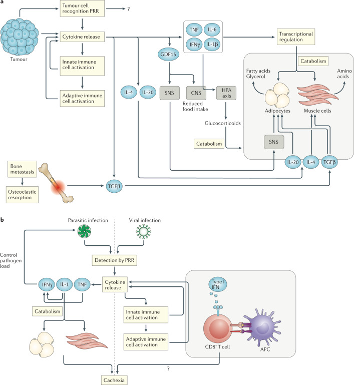
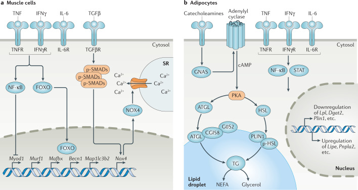
Diverse inflammatory diseases, infections and malignancies are associated with wasting syndromes. In many of these conditions, the standards for diagnosis and treatment are lacking due to our limited understanding of the causative molecular mechanisms. Here, we discuss the complex immunological context of cachexia, a systemic catabolic syndrome that depletes both fat and muscle mass with profound consequences for patient prognosis. We highlight the main cytokine and immune cell-driven pathways that have been linked to weight loss and tissue wasting in the context of cancer-associated and infection-associated cachexia. Moreover, we discuss the potential immunometabolic consequences of cachexia on the basis of newly identified pathways and explore the multilayered area of immunometabolic crosstalk both upstream and downstream of tissue catabolism. Collectively, this Review highlights the intricate relationship of the immune system with cachexia in the context of malignant and infectious diseases.


Conflict of interest statement

The authors declare no competing interests.

Figures

Fig. 1
Fig. 1. Relevant cytokines in the development of cachexia.
a | Tumorigenesis is associated with the release of a wide range of cytokines by tumour cells, by the surrounding tissue and from innate and adaptive immune cells downstream of pattern-recognition receptor (PRR) activation by tumour cell-associated damage-associated molecular patterns. Tumour necrosis factor (TNF), interferon-γ (IFNγ), IL-6 and IL-1β are able to induce tissue catabolism by modulating gene-expression profiles in both adipose tissue and muscle cells. IL-1β also contributes to cachexia through the central nervous system (CNS), where it modulates food intake and activates the hypothalamic–pituitary–adrenal (HPA) axis. The subsequent release of glucocorticoids contributes to tissue catabolism. GDF15 release induces weight loss through effects on the CNS that modulate food intake and by increasing adipose tissue lipolysis through sympathetic nervous system (SNS) signalling. IL-20 also contributes to adipose tissue depletion, while IL-4 shows protective effects on muscle cells. Transforming growth factor-β (TGFβ) released from tumour cells and/or bone (during bone metastasis-induced osteoclastic resorption) can induce adipose tissue fibrosis and compromise muscle strength. b | During parasitic infection, TNF and IL-1 release downstream of PRRs supports the production of IFNγ, which is important for controlling the pathogen load. IL-1 is also involved in the catabolism of adipose tissues and muscles. During viral infection, type I interferon (IFN) signalling to CD8+ T cells during antigen recognition and T cell activation is an important step in triggering cachexia.
Fig. 2
Fig. 2. Mechanisms of myocyte and adipocyte catabolism.
a | Cytokines induce muscle catabolism by regulating the transcription of various genes. NF-κB signalling suppresses Myod1 expression, which leads to inhibition of myoblast differentiation. Forkhead box protein O (FOXO) activation results in its translocation to the nucleus, where it upregulates the expression of genes involved in proteasomal degradation (for example, Murf1 and Mafbx) and autophagy (for example, Becn1 and Map1lc3b2, which encode beclin 1 and LC3B, respectively). Transforming growth factor-β (TGFβ)–SMAD signalling increases Nox4 expression, and NADPH oxidase 4 (NOX4) oxidizes and destabilizes the RYR1 calcium channel, leading to calcium leakage and compromised muscle contraction. b | Adipose tissue depletion is induced through the binding of catecholamines to β-adrenergic receptors, which activates cAMP phosphorylation of protein kinase A (PKA) downstream of GNAS and adenylyl cyclase. PKA is then able to phosphorylate adipose triglyceride lipase (ATGL) and hormone-sensitive lipase (HSL), resulting in their translocation to the surface of lipid droplets, where lipolysis occurs. ATGL-mediated lipolysis is modulated through its interaction with G0/G1 switch protein 2 (G0S2) and CGI58. Cytokine signalling in the adipose tissue induces transcriptional changes downstream of NF-κB and STAT signalling, resulting in an increase in the levels of lipolytic enzymes and a suppression of genes involved in lipogenesis. Perilipin 1 (PLIN1; encoded by Plin1) coats the surface of the lipid droplet and is also transcriptionally suppressed during lipolysis to expose a wider surface area to lipase activity. IFNγ, interferon-γ; IFNγR, interferon-γ receptor; IL-6R, IL-6 receptor; NEFA, non-esterified fatty acid; p-HSL, phosphorylated HSL; p-SMAD, phosphorylated SMAD; SR, sarcoplasmic reticulum; TG, triacylglycerol; TGFβR, TGFβ receptor; TNF, tumour necrosis factor; TNFR, tumour necrosis factor receptor.
Fig. 3
Fig. 3. Linking immune cells to cachexia.
A schematic summary of the immune cell types that correlate with cachexia in the setting of cancer or infection is shown. The mechanisms by which immune cells influence cachexia are not yet clear, and correlations do not necessarily imply causality between immune cell function and cachexia due to the entanglement of direct and/or indirect effects of the underlying disease and the cachectic processes. a | Experimental models of cancer-associated cachexia (CAC) demonstrate that an increased number of microglial cells in the brain has a protective effect against severe cachexia, potentially owing to microglial cell-mediated depletion of neutrophils, as high neutrophil numbers correlate with a higher degree of cachexia. b,c | In adipose tissue, macrophages protect against lipolysis, whereas in muscle tissue, macrophage numbers correlate with a higher degree of atrophy, and this is potentially affected by the macrophage polarization. In muscle, increased numbers of CD8+ T cells correlate negatively with cachexia, whereas the numbers of CD4+ T cells show both positive and negative correlations with cachexia, which is likely dependant on the specific subtype of T cell being measured in the population. d | The numbers of myeloid-derived suppressor cells (MDSCs) in the tumour, bone marrow and spleen positively correlate with cachexia. e | In parasite infection-associated cachexia, the numbers of CD4+FOXP3+ T cells negatively correlate with cachexia. f | During viral infection, antigen-specific CD8+ T cell responses show procachectic effects. Red and blue circles beside cell populations indicate procachetic or anticachectic effects of that population, respectively. IAC, infection-associated cachexia.

References

    1. Katz AM, Katz PB. Diseases of the heart in the works of Hippocrates. Br. Heart J. 1962;24:257–264. doi: 10.1136/hrt.24.3.257. - DOI - PMC - PubMed
    1. Slane J. Malarial cachexia in the Garo Hills. Ind. Med. Gaz. 1877;12:123–124. - PMC - PubMed
    1. Buchanan WJ. Case of general tuberculosis simulating malarial cachexia. Ind. Med. Gaz. 1898;33:139–140. - PMC - PubMed
    1. Baracos VE, Martin L, Korc M, Guttridge DC, Fearon KCHH. Cancer-associated cachexia. Nat. Rev. Dis. Primers. 2018;4:1–18. doi: 10.1038/nrdp.2017.105. - DOI - PubMed
    1. Keithley JK, Swanson B. HIV-associated wasting. J. Assoc. Nurses AIDS Care. 2013;24:S103–S111. doi: 10.1016/j.jana.2012.06.013. - DOI - PubMed

Publication types