Dynamic Oxygen Conditions Promote the Translocation of HIF-1 α to the Nucleus in Mouse Blastocysts
- PMID: 34608438
- PMCID: PMC8487385
- DOI: 10.1155/2021/5050527
Dynamic Oxygen Conditions Promote the Translocation of HIF-1 α to the Nucleus in Mouse Blastocysts
Abstract
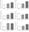
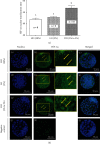
Oxygen tension is one of the most critical factors for mammalian embryo development and its survival. The HIF protein is an essential transcription factor that activated under hypoxic conditions. In this study, we evaluated the effect of dynamic oxygen conditions on the expression of embryonic genes and translocation of hypoxia-inducible factor-1α (HIF-1α) in cultured mouse blastocysts. Two-pronuclear (2PN) zygotes harvested from ICR mice were subjected to either high oxygen (HO; 20%), low oxygen (LO; 5%), or dynamic oxygen (DO; 5% to 2%) conditions. In the DO group, PN zygotes were cultured in 5% O2 from days 1 to 3 and then in 2% O2 till day 5 after hCG injection. On day 5, the percentage of blastocysts in the cultured embryos from each group was estimated, and the embryos were also subjected to immunocytochemical and gene expression analysis. We found that the percentage of blastocysts was similar among the experimental groups; however, the percentage of hatching blastocysts in the DO and LO groups was significantly higher than that in the HO group. The total cell number of blastocysts in the DO group was significantly higher than that of both the HO and LO groups. Further, gene expression analysis revealed that the expression of genes related to the embryonic development was significantly higher in the DO group than that in the HO and LO groups. Interestingly, HIF-1α mRNA expression did not significantly differ; however, HIF-1α protein translocation from the cytoplasm to the nucleus was significantly higher in the DO group than in the HO and LO groups. Our study suggests that dynamic oxygen concentrations increase the developmental capacity in mouse preimplantation embryos through activation of the potent transcription factor HIF-1α.
Copyright © 2021 Jungwon Choi et al.
Conflict of interest statement
The authors declare no conflicts of interest.
Figures


Similar articles
-
Effects of oxygen tension and IGF-I on HIF-1α protein expression in mouse blastocysts.J Assist Reprod Genet. 2013 Jan;30(1):99-105. doi: 10.1007/s10815-012-9902-z. Epub 2012 Dec 12. J Assist Reprod Genet. 2013. PMID: 23232974 Free PMC article.
-
Low oxygen tension increases mitochondrial membrane potential and enhances expression of antioxidant genes and implantation protein of mouse blastocyst cultured in vitro.J Ovarian Res. 2017 Jul 20;10(1):47. doi: 10.1186/s13048-017-0344-1. J Ovarian Res. 2017. PMID: 28728562 Free PMC article.
-
Oxygen-regulated gene expression in bovine blastocysts.Biol Reprod. 2004 Oct;71(4):1108-19. doi: 10.1095/biolreprod.104.028639. Epub 2004 May 26. Biol Reprod. 2004. PMID: 15163614
-
Involvement of Ca2+-independent phospholipase A2 in the translocation of hypoxia-inducible factor-1alpha to the nucleus under hypoxic conditions.Eur J Pharmacol. 2006 Nov 7;549(1-3):58-62. doi: 10.1016/j.ejphar.2006.08.026. Epub 2006 Aug 30. Eur J Pharmacol. 2006. PMID: 16979159
-
Single-embryo transcriptomic atlas of oxygen response reveals the critical role of HIF-1α in prompting embryonic zygotic genome activation.Redox Biol. 2024 Jun;72:103147. doi: 10.1016/j.redox.2024.103147. Epub 2024 Apr 2. Redox Biol. 2024. PMID: 38593632 Free PMC article.
Cited by
-
Hypoxia-Induced Changes in L-Cysteine Metabolism and Antioxidative Processes in Melanoma Cells.Biomolecules. 2023 Oct 7;13(10):1491. doi: 10.3390/biom13101491. Biomolecules. 2023. PMID: 37892173 Free PMC article.
-
Biphasic oxygen tension promotes the formation of transferable blastocysts in patients without euploid embryos in previous monophasic oxygen cycles.Sci Rep. 2023 Mar 15;13(1):4330. doi: 10.1038/s41598-023-31472-4. Sci Rep. 2023. PMID: 36922540 Free PMC article.
-
HIF1A contributes to the survival of aneuploid and mosaic pre-implantation embryos.bioRxiv [Preprint]. 2025 Apr 9:2023.09.04.556218. doi: 10.1101/2023.09.04.556218. bioRxiv. 2025. PMID: 39071426 Free PMC article. Preprint.
-
Embryos from Prepubertal Hyperglycemic Female Mice Respond Differentially to Oxygen Tension In Vitro.Cells. 2024 May 30;13(11):954. doi: 10.3390/cells13110954. Cells. 2024. PMID: 38891086 Free PMC article.
References
MeSH terms
Substances
LinkOut - more resources
Full Text Sources

