. 2022 Mar 12;24(3):429-441.
doi: 10.1093/neuonc/noab231.

The EGFRvIII transcriptome in glioblastoma: A meta-omics analysis

Affiliations

The EGFRvIII transcriptome in glioblastoma: A meta-omics analysis

Youri Hoogstrate et al. Neuro Oncol. .

Abstract

Background: EGFR is among the genes most frequently altered in glioblastoma, with exons 2-7 deletions (EGFRvIII) being among its most common genomic mutations. There are conflicting reports about its prognostic role and it remains unclear whether and how it differs in signaling compared with wildtype EGFR.

Methods: To better understand the oncogenic role of EGFRvIII, we leveraged 4 large datasets into 1 large glioblastoma transcriptome dataset (n = 741) alongside 81 whole-genome samples from 2 datasets.

Results: The EGFRvIII/EGFR expression ratios differ strongly between tumors and range from 1% to 95%. Interestingly, the slope of relative EGFRvIII expression is near-linear, which argues against a more positive selection pressure than EGFR wildtype. An absence of selection pressure is also suggested by the similar survival between EGFRvIII-positive and -negative glioblastoma patients. EGFRvIII levels are inversely correlated with pan-EGFR (all wildtype and mutant variants) expression, which indicates that EGFRvIII has a higher potency in downstream pathway activation. EGFRvIII-positive glioblastomas have a lower CDK4 or MDM2 amplification incidence than EGFRvIII-negative (P = .007), which may point toward crosstalk between these pathways. EGFRvIII-expressing tumors have an upregulation of "classical" subtype genes compared to those with EGFR-amplification only (P = 3.873e-6). Genomic breakpoints of the EGFRvIII deletions have a preference toward the 3'-end of the large intron-1. These preferred breakpoints preserve a cryptic exon resulting in a novel EGFRvIII variant and preserve an intronic enhancer.

Conclusions: These data provide deeper insights into the complex EGFRvIII biology and provide new insights for targeting EGFRvIII mutated tumors.

Keywords: EGFR; EGFRvIII; RNA-seq; breakpoints; glioblastoma.


Figures

Fig. 1
Fig. 1
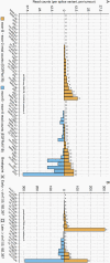
Range of EGFRvIII percentages relative to total EGFR. Results are split per dataset (top) and combined (bottom). Gray vertical lines (Intellance-2) indicate levels determined by both full and panel-based RNA-seq where the actual percentages reflect their mean. Mutation statuses are indicated underneath. N/A-values are indicated in black.
Fig. 2
Fig. 2
EGFRwt/EGFRvIII correlations. (A) EGFRwt and EGFRvIII correlation and (B) total EGFR and percentage of correlation, per dataset. (A) The correlations between EGFRwt and EGFRvIII are negative; (B) y-axis represents a surrogate for the total EGFR level (VST-transformed sum of EGFRwt + EGFRvIII junction-reads because the full gene EGFR read count is negatively affected by exons missing in EGFRvIII). Correlations are negative, indicating that tumors with higher proportions of EGFRvIII have lower levels of both variants combined.
Fig. 3
Fig. 3
(A) DE analysis between EGFR-amplified samples with (≥10%) and without EGFRvIII (<1%), with batch correction for the 4 datasets (Intellance-2, G-SAM, BELOB, and TCGA-GBM). 213/15.617 protein-coding genes were differentially expressed, including DLX1, DLX2, TSPAN31, TMPRSS7, PPBP, and DPT. Classical subtype genes are marked black. Overall LFCs were more often negative while the majority of the classical subtype genes had a positive LFC. (B) First two components of a supervised principal component analysis (213 DE genes). (C) Z-scores of Pearson correlation tests between genes and the relative EGFRvIII (x-axis) and EGFRwt (y-axis) levels, in samples with ≥10% EGFRvIII. Values near 0 represent no correlation, negative values represent a negative correlation, and positive values represent a positive correlation. Classical subtype genes are marked black. Genes with a significant difference (t test; q-value < 0.01) are marked purple. Genes showing a trend (q-value < 0.1) are marked blue.
Fig. 4
Fig. 4
Kaplan-Meier survival plots of patients with EGFR amplification with/without EGFRvIII. Patients included were from the BELOB trial, Intellance-2 TMZ/control arm, primary G-SAM tumors, and primary TCGA-GBM tumors. Difference in patient survival between EGFR-amplified glioblastoma patients with/without EGFRvIII (≥1% and ≥10%) was not significant and neither in each dataset separately (Supplementary Figure S7).
Fig. 5
Fig. 5
Overview of genomic EGFR locus (exons 1-11) and EGFRvIII breakpoints. From bottom to top: chr7, transcript annotations, late and early breakpoint regions, conservation (purple), H3K27ac intensity in GSC23 cells (green), and the actual breakpoints (blue and mustard). Genomic EGFRvIII breakpoints are indicated with mustard (RNA detected) and blue (DNA detected) bars on top.
Fig. 6
Fig. 6
Exon-B expression. Spliced read counts for exon-B (exon-B→exon-2: bars up and exon-B→exon-8: bars down) in tumors with RNA detected genomic EGFRvIII breakpoint. Tumors with a “late” intron-1 breakpoint (≥chr7:55.182.397) are marked with a square and “early” with a cross. Regular (A) and high (B) depth datasets were split. EGFRvIII exon-B variant reads (exon-B→exon-8) are only present in tumors with a late EGFRvIII breakpoint, which retains exon-B.

References

    1. Stupp R, Mason WP, van den Bent MJ, et al. ; European Organisation for Research and Treatment of Cancer Brain Tumor and Radiotherapy Groups; National Cancer Institute of Canada Clinical Trials Group . Radiotherapy plus concomitant and adjuvant temozolomide for glioblastoma. N Engl J Med. 2005;352(10):987–996. - PubMed
    1. McLendon R, Friedman A, Bigner D, et al. . Comprehensive genomic characterization defines human glioblastoma genes and core pathways. Nature. 2008;455(7216):1061–1068. - PMC - PubMed
    1. Parsons DW, Jones S, Zhang X, et al. . An integrated genomic analysis of human glioblastoma multiforme. Science. 2008;321(5897):1807–1812. - PMC - PubMed
    1. Brennan CW, Verhaak RGW, McKenna A, et al. . The somatic genomic landscape of glioblastoma. Cell. 2014;157(3):753. - PMC - PubMed
    1. French PJ, Eoli M, Sepulveda JM, et al. . Defining EGFR amplification status for clinical trial inclusion. Neuro Oncol. 2019;21(10):1263–1272. - PMC - PubMed

Publication types

Substances