

Proteomics changes after negative pressure wound therapy in diabetic foot ulcers
- PMID: 34608502
- PMCID: PMC8503750
- DOI: 10.3892/mmr.2021.12474
Proteomics changes after negative pressure wound therapy in diabetic foot ulcers
Abstract
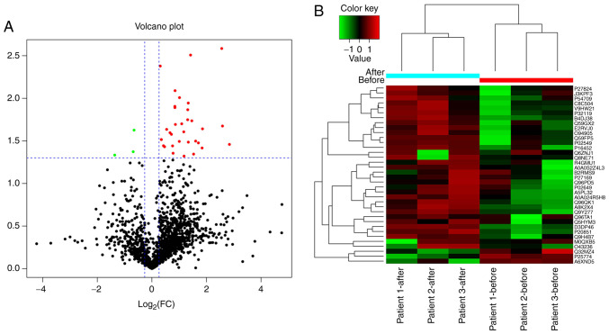
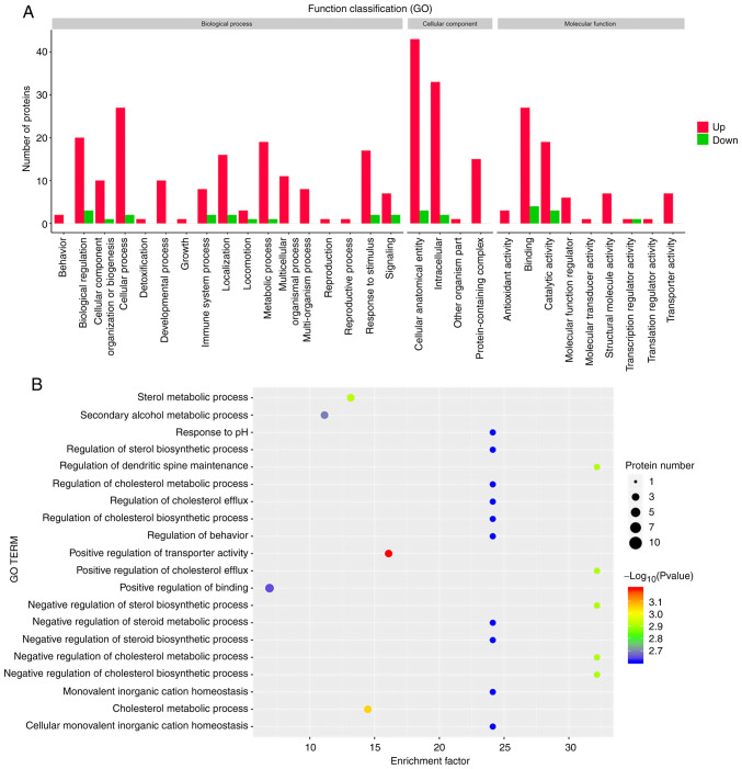
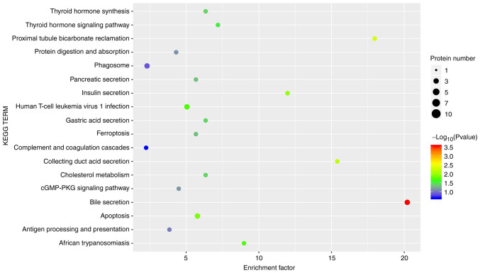
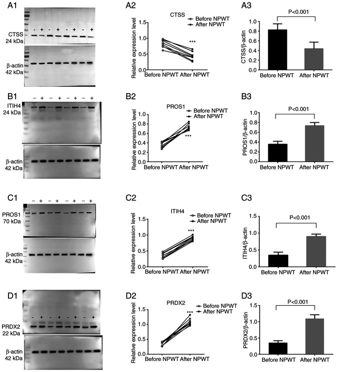
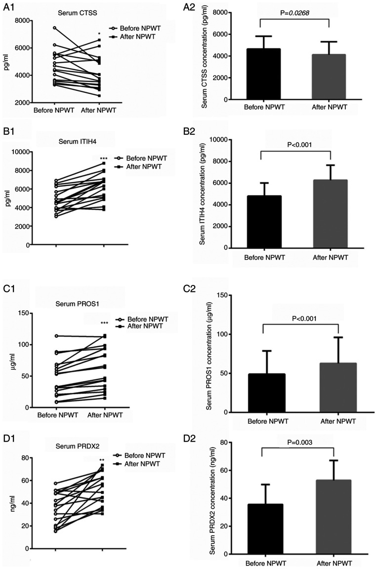
Label‑free quantitative mass spectrometry was used to analyze the differences in the granulation tissue protein expression profiles of patients with diabetic foot ulcers (DFUs) before and after negative‑pressure wound therapy (NPWT) to understand how NPWT promotes the healing of diabetic foot wounds. A total of three patients with DFUs hospitalized for Wagner grade 3 were enrolled. The patients received NPWT for one week. The granulation tissue samples of the patients prior to and following NPWT for one week were collected. The protein expression profiles were analyzed with label‑free quantitative mass spectrometry and the differentially expressed proteins (DEPs) in the DFU patients prior to and following NPWT for one week were identified. Gene Ontology and Kyoto Encyclopedia of Genes and Genomes analyses were conducted to annotate the DEPs and DEP‑associated signaling pathways. Western blotting and ELISA were performed to validate the results. By comparing the differences in the protein profiles of granulation tissue samples prior to and following NPWT for one week, 36 proteins with significant differences were identified (P<0.05); 33 of these proteins were upregulated and three proteins were downregulated. NPWT altered proteins mainly associated with antioxidation and detoxification, the cytoskeleton, regulation of the inflammatory response, complement and coagulation cascades and lipid metabolism. The functional validation of the DEPs demonstrated that the levels of cathepsin S in peripheral blood and granulation tissue were significantly lower than those prior to NPWT (P<0.05), while the levels of protein S isoform 1, inter α‑trypsin inhibitor heavy chain H4 and peroxiredoxin‑2 in peripheral blood and granulation tissue were significantly higher than those prior to NPWT (P<0.05). The present study identified multiple novel proteins altered by NPWT and laid a foundation for further studies investigating the mechanism of action of NPWT.
Keywords: cathepsin S; diabetic foot ulcer; differentially expressed proteins; inter α‑trypsin inhibitor heavy chain H4; label‑free quantitative mass spectrometry; negative‑pressure wound therapy; peroxiredoxin‑2; protein S isoform 1.
Conflict of interest statement
The authors declare that they have no competing interests.
Figures
Similar articles
-
Negative pressure wound therapy promotes wound healing by down-regulating miR-155 expression in granulation tissue of diabetic foot ulcers.Sci Rep. 2025 Feb 25;15(1):6733. doi: 10.1038/s41598-025-90643-7. Sci Rep. 2025. PMID: 40000694 Free PMC article. Clinical Trial.
-
Utility of 3D Wound Assessment in Monitoring Granulation Tissue Velocity Following Negative-Pressure Wound Therapy in Diabetic Foot Ulcers.Foot Ankle Int. 2025 Mar;46(3):287-294. doi: 10.1177/10711007251314805. Epub 2025 Feb 14. Foot Ankle Int. 2025. PMID: 39950603
-
Enhancing diabetic foot ulcer healing: Impact of the regulation of the FUS and ILF2 RNA‑binding proteins through negative pressure wound therapy.Int J Mol Med. 2024 Nov;54(5):103. doi: 10.3892/ijmm.2024.5427. Epub 2024 Sep 20. Int J Mol Med. 2024. PMID: 39301661 Free PMC article.
-
Negative pressure wound therapy is associated with up-regulation of bFGF and ERK1/2 in human diabetic foot wounds.Wound Repair Regen. 2014 Jul-Aug;22(4):548-54. doi: 10.1111/wrr.12195. Wound Repair Regen. 2014. PMID: 24809625 Review.
-
The Efficacy of Negative Pressure Wound Therapy (NPWT) on Healing of Diabetic Foot Ulcers: A Literature Review.Curr Diabetes Rev. 2024;20(8):1-11. doi: 10.2174/0115733998229877230926073555. Curr Diabetes Rev. 2024. PMID: 37921159 Review.
Cited by
-
What is slough? Defining the proteomic and microbial composition of slough and its implications for wound healing.Wound Repair Regen. 2024 Nov-Dec;32(6):783-798. doi: 10.1111/wrr.13170. Epub 2024 Apr 1. Wound Repair Regen. 2024. PMID: 38558438 Free PMC article.
-
Association between diabetic retinopathy and diabetic foot ulcer in patients with diabetes: A meta-analysis.Int Wound J. 2023 Dec;20(10):4077-4082. doi: 10.1111/iwj.14299. Epub 2023 Aug 9. Int Wound J. 2023. Retraction in: Int Wound J. 2025 Mar;22(3):e70312. doi: 10.1111/iwj.70312. PMID: 37554103 Free PMC article. Retracted.
-
Key extracellular proteins and TF-miRNA co-regulatory network in diabetic foot ulcer: Bioinformatics and experimental insights.PLoS One. 2024 Jul 22;19(7):e0307205. doi: 10.1371/journal.pone.0307205. eCollection 2024. PLoS One. 2024. PMID: 39037979 Free PMC article.
-
A bibliometric analysis of global research hotspots and development trends in diabetic wound treatment.Front Clin Diabetes Healthc. 2025 Jun 20;6:1603206. doi: 10.3389/fcdhc.2025.1603206. eCollection 2025. Front Clin Diabetes Healthc. 2025. PMID: 40620799 Free PMC article.
-
Negative pressure wound therapy promotes wound healing of diabetic foot ulcers by up-regulating PRDX2 in wound margin tissue.Sci Rep. 2023 Sep 27;13(1):16192. doi: 10.1038/s41598-023-42634-9. Sci Rep. 2023. PMID: 37758743 Free PMC article.
References
MeSH terms
Substances
LinkOut - more resources
Full Text Sources
Medical
Research Materials
Miscellaneous
