. 2021 Oct 5;187(2):480-484.
doi: 10.1093/plphys/kiab306.

The latest HyPe(r) in plant H2O2 biosensing

Affiliations

The latest HyPe(r) in plant H2O2 biosensing

José Manuel Ugalde et al. Plant Physiol. .

Abstract

HyPer7 senses minute amounts of H2O2 independent of pH and the glutathione redox potential and enables detection of physiological H2O2 fluxes within the cytosol and between subcellular compartments.


Figures
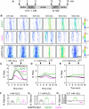
Figure 1
Figure 1
Oxidation and reduction of different genetically encoded H2O2 probes in the cytosol of wild-type Arabidopsis seedlings. A, Experimental design for sequential perfusion with different concentrations of H2O2, imaging buffer and 10 mM DTT. B, Confocal microscopy images of root cells from 7-d-old seedlings expressing HyPer7, HyPer, or roGFP2-Orp1, all targeted to the cytosol. The false color ratio images show the fluorescence ratios calculated from two separate images collected with excitation at 488 and 405 nm. Emission was collected at 508–535 nm. Ratios are 488/405 nm for HyPer7 and HyPer, and 405/488 nm for roGFP2-Orp1. Bar, 50 μm. C–E, Typical time courses showing the dynamic response of the probes to transient oxidation and subsequent reduction by the indicated treatments (arrows). All ratios are normalized to the starting values at t = 0 min. n = 2–4 replicates. F–H, Slopes of the ratio value changes after H2O2 perfusion in the indicated concentrations. The slopes were calculated from ratio values between 3.9 and 4.7 min. Data indicate the mean values ± sd. n = 2–4 replicates.
Figure 2
Figure 2
Laser illumination of green tissues during scanning is sufficient to produce release of H2O2 from chloroplasts. A and B, Laser-induced oxidation is more visible in cells expressing HyPer7 compared with cells with HyPer or roGFP2-Orp1. Plants expressing cpYFP were included to test for putative concomitant pH changes. The false color ratio images show the fluorescence ratios calculated from two separate images collected with excitation at 488 and 405 nm. Emission was collected at 508–535 nm. Ratios are 488/405 nm for HyPer7, HyPer, and cpYFP, and 405/488 nm for roGFP2-Orp1. Bar, 20 μm. C, Ratio values of purified recombinant protein imaged with the same instruments settings as in (A). The protein was reduced with TCEP prior to the measurement. D and E, Inhibition of photosynthetic electron transport blocks laser induced oxidation of HyPer7 in the cytosol. Seedlings were incubated in imaging buffer supplemented with 20 µM DCMU for 45 min in the dark prior to the measurement. Bar, 20 μm. F–H, Laser-induced oxidation of HyPer7 in a defined region of interest (magenta square) is reversible. Letters indicate time windows for laser-induced oxidation caused by high-frequency imaging at high zoom (a–b and c–d) and a recovery phase with intermittent imaging at low zoom (b–c and d–e). G, Fluorescence measured from the two independent channels (405 and 488 nm), normalized to the value at t = 0 min. H, 488/405 nm ratio for the values shown in panel G. All ratios in B, C, E, and H are normalized to the ratio value at t = 0 min. Ratios are means + sd, n = 4–11 replicates.

Similar articles

Cited by

References

    1. Belousov VV, Fradkov AF, Lukyanov KA, Staroverov DB, Shakhbazov KS, Terskikh AV, Lukyanov S (2006) Genetically encoded fluorescent indicator for intracellular hydrogen peroxide. Nat Methods 3: 281–286 - PubMed
    1. Bilan DS, Belousov VV (2016) HyPer family probes: State of the art. Antioxid Redox Signal 24: 731–751 - PubMed
    1. Costa A, Drago I, Behera S, Zottini M, Pizzo P, Schroeder JI, Pozzan T, Lo Schiavo F (2010) H2O2 in plant peroxisomes: An in vivo analysis uncovers a Ca2+-dependent scavenging system. Plant J 62: 760–772 - PMC - PubMed
    1. Exposito-Rodriguez M, Laissue PP, Yvon-Durocher G, Smirnoff N, Mullineaux PM (2017) Photosynthesis-dependent H2O2 transfer from chloroplasts to nuclei provides a high-light signalling mechanism. Nat Commun 8: 49. - PMC - PubMed
    1. Fichman Y, Miller G, Mittler R (2019) Whole-plant live imaging of reactive oxygen species. Mol Plant 12: 1203–1210 - PubMed

Publication types

MeSH terms

Substances