. 2021 Jan-Mar;62(1):133-149.
doi: 10.47162/RJME.62.1.13.

Immunohistochemical evaluation of D2-40, Galectin-3, Maspin and MCM7 expression in palate squamous cell carcinomas

Affiliations

Immunohistochemical evaluation of D2-40, Galectin-3, Maspin and MCM7 expression in palate squamous cell carcinomas

Adrian Pătru et al. Rom J Morphol Embryol. 2021 Jan-Mar.

Abstract

Squamous cell carcinoma (SCC) is the most frequent cancer in oral cavity and its prognosis has exhibited little improvement in the last decades. Although much less common palate SCCs manifests a higher local aggression invading very quickly the adjacent muscles and jawbones, thus being able frequently to lead to dysfunctions in chewing, swallowing, and speech. To elucidate what underlies such local aggression, we investigated the immunohistochemical expression in palate SCCs of Podoplanin (D2-40), Galectin-3 (Gal-3), mammary serine protease inhibitor (Maspin) and minichromosome maintenance complex component 7 (MCM7), markers that are known to be involved in tumor invasiveness. We found a progressive increase in reactivity for D2-40 and MCM7 from the normal epithelium toward dysplastic epithelium and respectively to SCC, which suggests the intervention of these markers in the early stages of squamous cell carcinogenesis in the palate. The highest D2-40, Gal-3 and MCM7 reactivity was observed in basaloid and in poorly differentiated (G3) palate SCCs, while for Maspin the well-differentiated (G1) palate SCCs were the most reactive. The first three markers mentioned above were most intensely expressed at the invasion front, while the Maspin reactivity was low or absent at this level. Statistically, we found significant stratification on localization, grading, muscle invasion, and survival for all investigated markers, but with very high direct correlations between D2-40, Gal-3, and MCM7 immunoreactive score (IRS) values, while between the Maspin and each of the previous markers there were very high inverse correlations. Overall, all these investigate markers proved to be responsible for the local invasiveness and regional lymph node metastasis, thus allowing a prognostic and therapeutic stratification of patients with palate SCCs.


Conflict of interest statement

The authors declare that there is no conflict of interests regarding the publication of this paper. All authors read and approved the final manuscript.

Figures
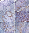
Figure 1
Figure 1
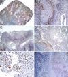
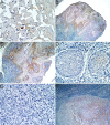
D2-40 immunoreactivity: (A) At the level of basal layer from normal epithelium adjacent to the tumors with cytoplasmic and membrane pattern; (B) The reactivity extended to the suprabasal layer in the hyperplastic and dysplastic epithelium; (C) Membranous reactivity in the lymphatic vessels both from tumor and normal samples; (D) The highest reactivity was noticed in the poorly differentiated (G3) palate SCC subtype; (E) This was followed by the basaloid variant, especially at the periphery of tumor proliferations; (F) A similar reactivity was observed in moderately differentiated (G2) palate SCC subtype, more obvious at the invasive front. Anti-D2-40 antibody immunolabeling: (A–F) ×200. SCC: Squamous cell carcinoma
Figure 2
Figure 2
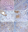
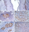
D2-40 immunoreactivity: (A) An even lower reactivity was recorded in papillary palate SCC cases; (B) These were followed by the well-differentiated (G1) palate SCC cases where the reactivity was restricted to the tumor cells from the periphery of neoplastic proliferation, with a dominant membranous subcellular pattern; (C) The lowest reactivity was recorded in the verrucous subtype; (D) This reactivity was similar to that of acantholytic SCC cases; (E) In the acantholytic cases, the D2-40 reactivity was also present in some of the discohesive tumor cells, with both cytoplasmic and membranous subcellular patterns; (F) A lower reactivity was also recorded in the metastatic neoplastic proliferations from loco-regional lymph nodes. Anti-D2-40 antibody immunolabeling: (A, C and D) ×50; (B) ×200; (E) ×400; (F) ×100. SCC: Squamous cell carcinoma
Figure 3
Figure 3
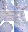
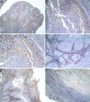
Gal-3 immunoreactivity: (A) Weak nuclear reactivity in basal and parabasal layers, and with cytoplasmic pattern in the superficial layers of normal epithelium; (B) Intense reaction in the serous acini and ductal luminal cells of minor salivary glands with a mixed cytoplasmic, membranous, and nuclear pattern – also, a moderate reactivity was noticed in the nerve fibers with cytoplasmic and membranous pattern; (C) A strong cytoplasmic and membranous immunostaining at the level of the excretory units from minor salivary glands; (D) Maximum reactivity was observed in the basaloid palate SCC cases with prevalent nuclear pattern; (E) A slightly lower reactivity was observed in the G3 palate SCCs, which has a cytoplasmic dominant pattern, but with more intense reaction at the nuclear level; (F) A similar reactivity was present in the G2 tumors. Anti-Gal-3 antibody immunolabeling: (A–D, and F) ×200; (E) ×400. G2: Moderately differentiated tumor; G3: Poorly differentiated tumor; Gal-3: Galectin-3; SCC: Squamous cell carcinoma
Figure 4
Figure 4
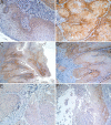
Gal-3 immunoreactivity: (A) In the acantholytic variant, the cytoplasmic pattern was the dominant one, this reactivity decreasing progressively towards the lumens; (B) In the lumens, some tumor acantholytic cells retained the reactivity; (C) A lower reactivity was recorded in G1 palate SCCs, with a prevalent cytoplasmic subcellular pattern, followed by the membranous one; (D) The lowest Gal-3 reactivity was observed in the verrucous subtype; (E) This was followed by the papillary palate SCC that had a similar pattern as was noticed in G1 palate SCCs; (F) A lower cytoplasmic reactivity was also noticed in metastatic tumor proliferations from the loco-regional lymph nodes. Anti-Gal-3 antibody immunolabeling: (A and F) ×100; (B and C) ×200; (D and E) ×50. G1: Well-differentiated tumor; Gal-3: Galectin-3; SCC: Squamous cell carcinoma
Figure 5
Figure 5
Maspin immunoreactivity: (A) A moderate cytoplasmic reactivity especially in the basal and suprabasal layers and also some diffuse nuclear reactivity in keratinocytes from the thickness of the normal epithelium; (B) G1 palate SCCs were the most reactive, generally with a dominant cytoplasmic and membranous pattern but also focally with a nuclear pattern; (C) The verrucous case was less reactive; (D) These were followed by the G2 palate SCCs, with similar immunostaining pattern as was observed in G1 palate SCCs; (E) In these cases was obvious a decrease in reactivity to the invasion front; (F) A similar reactivity was noticed in the acantholytic carcinomas, with an obvious decrease in reactivity in acantholysis areas. Anti-Maspin antibody immunolabeling: (A, B, and E) ×200; (C) ×50; (D and F) ×100. G1: Well-differentiated tumor; G2: Moderately differentiated tumor; Maspin: Mammary serine protease inhibitor; SCC: Squamous cell carcinoma
Figure 6
Figure 6
Maspin immunoreactivity: (A) In the acantholysis areas, only a few acantholytic cells have retained Maspin reactivity; (B) The papillary SCCs had a similar immunostaining pattern as was observed in G2 palate SCCs; (C) The highest reactivity was observed in cells from the papillae periphery; (D) In one basaloid palate SCC, a weak cytoplasmic reactivity was observed in neoplastic cells from the middle of tumor proliferations; (E) No Maspin reactivity was noticed in all G3 palate SCCs; (F) A weak Maspin cytoplasmic reactivity was observed at the level of metastatic neoplastic proliferations from lymph nodes metastases. Anti-Maspin antibody immunolabeling: (A, D, and E) ×400; (B) ×50; (C and F) ×100. G2: Moderately differentiated tumor; G3: Poorly differentiated tumor; Maspin: Mammary serine protease inhibitor; SCC: Squamous cell carcinoma
Figure 7
Figure 7
MCM7 immunoreactivity: (A) In the normal epithelium from tumor samples, the MCM7 reactivity was limited to the basal and parabasal layers heaving a nuclear pattern; (B) In the associated hyperplastic and dysplastic lesions, the reactivity increase towards the middle of the epithelium; (C) A lower nuclear reactivity was observed in some glandular cells from minor salivary gland and a high intensity in the germinal center cells from lymphoid follicles; (D) The highest MCM7 reactivity was recorded in G3 palate SCCs, with an exclusively nuclear pattern; (E) A similar immunostaining pattern was present in the basaloid subtype, the reactivity being less obvious towards the center of the tumor islands; (F) Then followed the G2 palate SCCs, with the highest reactivity in the cells from the periphery of tumor proliferations, the intensity of the immunostaining decreasing progressively inwards directly proportional to the degree of keratinization. Anti-MCM7 antibody immunolabeling: (A–E) ×200; (F) ×100. G2: Moderately differentiated tumor; G3: Poorly differentiated tumor; MCM7: Minichromosome maintenance complex component 7; SCC: Squamous cell carcinoma
Figure 8
Figure 8
MCM7 immunoreactivity: (A) A slightly lower reactivity was recorded in both papillary palate SCCs; (B) In the acantholytic palate SCCs, the nuclear reactivity was present in the peripheral layers of acantholytic tumor islands; (C) Particularly, in this variant of palate SCC there was a cytoplasmic reactivity in acantholytic cells; (D) In the G1 palate SCCs, the high intensity nuclear immunostaining was noticed in peripheral layers and no reactivity at the level of keratin pearls; (E) A similar reactivity was also noticed in the palate SCC verrucous case; (F) A nuclear immunostaining reactivity was also recorded in the metastatic neoplastic proliferations from lymph nodes metastases. Anti-MCM7 antibody immunolabeling: (A, E and F) ×50; (B and D) ×100; (C) ×200. G1: Well-differentiated tumor; MCM7: Minichromosome maintenance complex component 7; SCC: Squamous cell carcinoma
Figure 9
Figure 9
IRS values distribution on localization for all investigated markers. D2-40, Gal-3, MCM7 show a higher frequency of Class B (higher) score in the soft palate, while Maspin shows a higher frequency of Class A score. Class A include the IRS values ranging from 1 to 6; Class B comprise the IRS values higher than 6. D2-40: Podoplanin; Gal-3: Galectin-3; IRS: Immunoreactive score; Maspin: Mammary serine protease inhibitor; MCM7: Minichromosome maintenance complex component 7
Figure 10
Figure 10
IRS values distribution on tumor grading for all investigated markers. D2-40, Gal-3, and MCM7 show predominant Class A scores in G1, combined Class A and B in G2 and predominant Class B in G3, while Maspin scores are mirrored. Class A include the IRS values ranging from 1 to 6; Class B comprise the IRS values higher than 6. D2-40: Podoplanin; G1: Well-differentiated tumor; G2: Moderately differentiated tumor; G3: Poorly differentiated tumor; Gal-3: Galectin-3; IRS: Immunoreactive score; Maspin: Mammary serine protease inhibitor; MCM7: Minichromosome maintenance complex component 7
Figure 11
Figure 11
IRS values distribution on T stage for all investigated markers. D2-40, Gal-3, and MCM7 show a similar distribution with a concentration of Class A scores in T1 and T2, and concentration of Class B scores in T3 and T4. Again, Maspin has a mirrored aspect with T1 and T2 concentration scores from Class B, and T3 and T4 from Class A. Class A include the IRS values ranging from 1 to 6; Class B comprise the IRS values higher than 6. D2-40: Podoplanin; Gal-3: Galectin-3; IRS: Immunoreactive score; Maspin: Mammary serine protease inhibitor; MCM7: Minichromosome maintenance complex component 7

Similar articles

Cited by

References

    1. Bray F, Ferlay J, Soerjomataram I, Siegel RL, Torre LA, Jemal A. Global cancer statistics 2018: GLOBOCAN estimates of incidence and mortality worldwide for 36 cancers in 185 countries. CA Cancer J Clin. 2018;68(6):394–424. - PubMed
    1. Kim JW, Park Y, Roh JL, Cho KJ, Choi SH, Nam SY, Kim SY. Prognostic value of glucosylceramide synthase and P-glycoprotein expression in oral cavity cancer. Int J Clin Oncol. 2016;21(5):883–889. - PubMed
    1. Farhood Z, Simpson M, Ward GM, Walker RJ, Osazuwa-Peters N. Does anatomic subsite influence oral cavity cancer mortality? A SEER database analysis. Laryngoscope. 2019;129(6):1400–1406. - PubMed
    1. Brierley JD, Gospodarowicz MK, Wittekind C, editors. TNM classification of malignant tumors. 8. Union for International Cancer Control (UICC) Willey-Blackwell John Wiley & Sons Ltd; 2017. pp. 272–272.
    1. Urken ML. Advances in head and neck reconstruction. Laryngoscope. 2003;113(9):1473–1476. - PubMed