Comparing PRP and bone marrow aspirate effects on cartilage defects associated with partial meniscectomy: a confocal microscopy study on animal model
- PMID: 34609430
- PMCID: PMC8597375
- DOI: 10.47162/RJME.62.1.27
Comparing PRP and bone marrow aspirate effects on cartilage defects associated with partial meniscectomy: a confocal microscopy study on animal model
Abstract
Aim: The aim of our study was to assess the therapeutic effects of platelet-rich plasma (PRP) and bone marrow aspirate concentrate (BMAC) in an animal knee lesion complex associating a large osteochondral defect and meniscal defect resulted from partial meniscectomy, a clinical situation that occurs quite often in orthopedic practice.
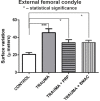
Materials and methods: Twenty-one male rabbits were included in the study, and all underwent initial surgery on the right knee to create the osteochondral defect on the internal femoral condyle, and remove the anterior horn of the internal meniscus, simulating a clinical situation. Rabbits were separated in three study groups: control, PRP group, in which three PRP injections were administered, and BMAC group, in which one single BMAC injection was administered. At the end of the six months follow-up period, knees were harvested and further analyzed using confocal microscopy and three-dimensional (3D) reconstruction of the articular surface.
Results: Therapeutic groups had better results concerning articular surface remodeling and joint degeneration indicators in comparison to trauma group.
Conclusions: Our results suggest that using post-operative regenerative therapies does improve final results concerning surface contact remodeling that was investigated using confocal microscopy and should be considered a valid treatment adjuvant in managing patients with this type of lesion complex, as it improves global joint outcome.
Conflict of interest statement
The authors declare that they have no conflict of interests.
Figures


Similar articles
-
Magnetic resonance imaging combined with histological evaluation of repair process using the microfracture technique in an association of osteocartilaginous and meniscal surgically induced lesions of the knee. In vivo experiment on a rabbit model.Rom J Morphol Embryol. 2024 Jan-Mar;65(1):89-97. doi: 10.47162/RJME.65.1.11. Rom J Morphol Embryol. 2024. PMID: 38527988 Free PMC article.
-
KNEE JOINT STRUCTURAL CHANGES IN OSTEOARTHRITIS AND INJECTIONS OF PLATELET RICH PLASMA AND BONE MARROW ASPIRATE CONCENTRATE.Georgian Med News. 2020 Jun;(303):184-188. Georgian Med News. 2020. PMID: 32841203
-
Bone Marrow Aspirate Concentrate versus Platelet Rich Plasma or Hyaluronic Acid for the Treatment of Knee Osteoarthritis.Medicina (Kaunas). 2021 Nov 2;57(11):1193. doi: 10.3390/medicina57111193. Medicina (Kaunas). 2021. PMID: 34833411 Free PMC article. Clinical Trial.
-
PRP and BMAC for Musculoskeletal Conditions via Biomaterial Carriers.Int J Mol Sci. 2019 Oct 25;20(21):5328. doi: 10.3390/ijms20215328. Int J Mol Sci. 2019. PMID: 31717698 Free PMC article. Review.
-
The Role of Bone Marrow Aspirate Concentrate for the Treatment of Focal Chondral Lesions of the Knee: A Systematic Review and Critical Analysis of Animal and Clinical Studies.Arthroscopy. 2019 Jun;35(6):1860-1877. doi: 10.1016/j.arthro.2018.11.073. Epub 2019 Mar 11. Arthroscopy. 2019. PMID: 30871903
Cited by
-
Evaluating Neutrophil Gelatinase-Associated Lipocalin in Pediatric CKD: Correlations with Renal Function and Mineral Metabolism.Pediatr Rep. 2024 Dec 9;16(4):1099-1114. doi: 10.3390/pediatric16040094. Pediatr Rep. 2024. PMID: 39728735 Free PMC article.
-
Assessing Differential Transfusion Requirements for Children with Congenital Malformations vs. Pediatric Acute Abdomen Emergencies.Diagnostics (Basel). 2024 Oct 4;14(19):2216. doi: 10.3390/diagnostics14192216. Diagnostics (Basel). 2024. PMID: 39410620 Free PMC article.
-
Comparative Evaluation of Temporomandibular Disorders and Dental Wear in Video Game Players.J Clin Med. 2024 Dec 25;14(1):31. doi: 10.3390/jcm14010031. J Clin Med. 2024. PMID: 39797113 Free PMC article.
-
Magnetic resonance imaging combined with histological evaluation of repair process using the microfracture technique in an association of osteocartilaginous and meniscal surgically induced lesions of the knee. In vivo experiment on a rabbit model.Rom J Morphol Embryol. 2024 Jan-Mar;65(1):89-97. doi: 10.47162/RJME.65.1.11. Rom J Morphol Embryol. 2024. PMID: 38527988 Free PMC article.
-
A Pre-clinical Standard Operating Procedure for Evaluating Orthobiologics in an In Vivo Rat Spinal Fusion Model.J Orthop Sports Med. 2022;4(3):224-240. doi: 10.26502/josm.511500060. Epub 2022 Sep 5. J Orthop Sports Med. 2022. PMID: 36203492 Free PMC article.
References
-
- Willers C, Wood DJ, Zheng MH. A current review on the biology and treatment of the articular cartilage defects (part I & part II) J Musculoskelet Res. 2003;7(3&4):157–181.
-
- Curl WW, Krome J, Gordon ES, Rushing J, Smith BP, Poehling GG. Cartilage injuries: a review of 31,516 knee arthroscopies. Arthroscopy. 1997;13(4):456–460. - PubMed
-
- Browne JE, Branch TP. Surgical alternatives for treatment of articular cartilage lesions. J Am Acad Orthop Surg. 2000;8(3):180–189. - PubMed
-
- Akeda K, An HS, Okuma M, Attawia M, Miyamoto K, Thonar EJMA, Lenz ME, Sah RL, Masuda K. Platelet-rich plasma stimulates porcine articular chondrocyte proliferation and matrix biosynthesis. Osteoarthritis Cartilage. 2006;14(12):1272–1280. - PubMed
-
- Krüger JP, Hondke S, Endres M, Pruss A, Siclari A, Kaps C. Human platelet-rich plasma stimulates migration and chondrogenic differentiation of human subchondral progenitor cells. J Orthop Res. 2012;30(6):845–852. - PubMed
MeSH terms
LinkOut - more resources
Full Text Sources
Research Materials

