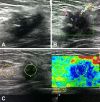
Analysis of breast cancer subtypes and their correlations with receptors and ultrasound
- PMID: 34609431
- PMCID: PMC8597389
- DOI: 10.47162/RJME.62.1.28
Analysis of breast cancer subtypes and their correlations with receptors and ultrasound
Abstract
The study aim was to evaluate the ultrasound (US) signs of the mammary lesions classified in the Breast Imaging-Reporting and Data System (BI-RADS) score category 3, 4, and 5, corresponding to US BI-RADS. It also followed the correlation between US changes of lesions suggestive for malignancy with the histopathological results and evaluated the proper management of those lesions. There were correlations of breast cancer (BC) subtypes with the receptors [estrogen receptor (ER), progesterone receptor (PR), human epidermal growth factor receptor 2 (HER2)], and Ki67 index, and the signs of conventional ultrasonography and US elastography. We selected 108 female patients examined with US, mammography and fine-needle biopsy who presented suspicions for malignancy lesions. Following the immunohistochemical analysis, they were classified in one of the BC subtypes. According to chi-squared analysis of molecular cancer subtypes correlation to receptors and Ki67 index, we found significant associations between both luminal A and luminal B HER2-negative subtypes and hormone receptors (ER, PR). These have an inverse relationship with Ki67 index elevated values; luminal B HER2-positive subtype has a direct association with HER2 presence; HER2-enriched subtype was statistically significant associated to HER2 presence and elevated Ki67 index values but had an inverse relationship to hormone receptors (ER, PR); triple-negative subtype was strongly associated to Ki67 index values and inversely correlated to ER and PR. We found luminal A subtype as being the most common and luminal B HER2-positive subtype as having the fewer cases.
Conflict of interest statement
The authors declare that they have no conflict of interests.
Figures


References
-
- Bangal VB, Shinde KK, Gavhane SP, Singh RK. Breast carcinoma in women - a rising threat. Int J Biomed Adv Res. 2013;4(2):73–76.
-
- Wen X, Yu Y, Yu X, Cheng W, Wang Z, Liu L, Zhang L, Qin L, Tian J. Correlations between ultrasonographic findings of invasive lobular carcinoma of the breast and intrinsic subtypes. Ultraschall Med. 2019;40(6):764–770. - PubMed
-
- Gheonea IA, Donoiu L, Camen D, Popescu FC, Bondari S. Sonoelastography of breast lesions: a prospective study of 215 cases with histopathological correlation. Rom J Morphol Embryol. 2011;52(4):1209–1214. - PubMed
MeSH terms
Substances
LinkOut - more resources
Full Text Sources
Medical
Research Materials
Miscellaneous

