. 2021 Dec;95(12):3803-3813.
doi: 10.1007/s00204-021-03164-3. Epub 2021 Oct 5.

Cell survival after DNA damage in the comet assay

Affiliations

Cell survival after DNA damage in the comet assay

Ezgi Eyluel Bankoglu et al. Arch Toxicol. 2021 Dec.

Abstract

The comet assay is widely used in basic research, genotoxicity testing, and human biomonitoring. However, interpretation of the comet assay data might benefit from a better understanding of the future fate of a cell with DNA damage. DNA damage is in principle repairable, or if extensive, can lead to cell death. Here, we have correlated the maximally induced DNA damage with three test substances in TK6 cells with the survival of the cells. For this, we selected hydrogen peroxide (H2O2) as an oxidizing agent, methyl methanesulfonate (MMS) as an alkylating agent and etoposide as a topoisomerase II inhibitor. We measured cell viability, cell proliferation, apoptosis, and micronucleus frequency on the following day, in the same cell culture, which had been analyzed in the comet assay. After treatment, a concentration dependent increase in DNA damage and in the percentage of non-vital and apoptotic cells was found for each substance. Values greater than 20-30% DNA in tail caused the death of more than 50% of the cells, with etoposide causing slightly more cell death than H2O2 or MMS. Despite that, cells seemed to repair of at least some DNA damage within few hours after substance removal. Overall, the reduction of DNA damage over time is due to both DNA repair and death of heavily damaged cells. We recommend that in experiments with induction of DNA damage of more than 20% DNA in tail, survival data for the cells are provided.

Keywords: Cell death and comet assay; DNA damage; DNA repair.


Conflict of interest statement

The authors declare that they have no conflict of interest.

Figures

Fig. 1
Fig. 1
DNA strand breaks induced by 60 µM H2O2 over time in alkaline comet assay. *p ≤ 0.05 vs. Control at the same time point
Fig. 2
Fig. 2
DNA strand breaks induced by 200 µM MMS over time in alkaline comet assay. *p ≤ 0.05 vs. Control at the same time point
Fig. 3
Fig. 3
DNA strand breaks induced by 1 µM Etoposide over time in alkaline comet assay. *p ≤ 0.05 vs. Control at the same time point
Fig. 4
Fig. 4
Concentration dependent increase in DNA damage after 0.5 h H2O2 treatment. *p ≤ 0.05 vs. 0 µM H2O2
Fig. 5
Fig. 5
Concentration dependent increase in DNA damage after 4 h MMS treatment. *p ≤ 0.05 vs. 0 µM MMS
Fig. 6
Fig. 6
Concentration dependent increase in DNA damage after 4 h etoposide treatment. *p ≤ 0.05 vs. 0 µM etoposide
Fig. 7
Fig. 7
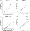
Correlation between the percentage of lost/apoptotic cells and the percentage of DNA in tail. (A) Correlation graph for H2O2 treatment. (B) Correlation graph for MMS treatment. (C) Correlation graph for etoposide treatment. (D) Correlation graphic for all three substances. Spearman correlation analysis was performed for each substance and the results showed a significant positive correlation between the percentage of lost/apoptotic cells and the percentage of DNA in tail
Fig. 8
Fig. 8
DNA damage reduction after H2O2 treatment over time in the comet assay. Treatment duration was 0.5 h and DNA damage was measured after medium change. *p ≤ 0.05 vs. Control at the same time point and ∆ ≤ 0.05 vs. 60 µM H2O2 at 0 h
Fig. 9
Fig. 9
DNA damage reduction after MMS treatment over time in the comet assay. Treatment duration was 4 h and DNA damage was measured after the medium change. *p ≤ 0.05 vs. Control at the same time point and ∆ ≤ 0.05 vs. 200 µM MMS at 0 h
Fig. 10
Fig. 10
DNA damage reduction after etoposide treatment over time in the comet assay. Treatment duration was 4 h and DNA damage was measured after the medium change. *p ≤ 0.05 vs. Control at the same time point and ∆ ≤ 0.05 vs. 1 µM etoposide at 0 h

References

    1. Anderson D, Dhawan A, Laubenthal J. The comet assay in human biomonitoring. In: Dhawan A, Bajpayee M, editors. Genotoxicity assessment: methods and protocols. Springer; 2019. pp. 259–274. - PubMed
    1. Azqueta A, Slyskova J, Langie SAS, O’Neill Gaivão I, Collins A. Comet assay to measure DNA repair: approach and applications. Front Genet. 2014 doi: 10.3389/fgene.2014.00288. - DOI - PMC - PubMed
    1. Azqueta A, Langie SAS, Boutet-Robinet E, et al. DNA repair as a human biomonitoring tool: comet assay approaches. Mutation Res Rev Mutation Res. 2019;781:71–87. doi: 10.1016/j.mrrev.2019.03.002. - DOI - PubMed
    1. Azqueta A, Ladeira C, Giovannelli L, et al. Application of the comet assay in human biomonitoring: an hCOMET perspective. Mutat Res. 2020;783:108288. doi: 10.1016/j.mrrev.2019.108288. - DOI - PubMed
    1. Baldwin EL, Osheroff N. Etoposide, topoisomerase II and cancer. Curr Med Chem Anticancer Agents. 2005;5(4):363–372. doi: 10.2174/1568011054222364. - DOI - PubMed

MeSH terms

LinkOut - more resources