Review
. 2022 Jan 1;95(1129):20210538.
doi: 10.1259/bjr.20210538. Epub 2021 Oct 5.

Comprehensive review of the recent advances in devices for endovascular treatment of complex brain aneurysms

Affiliations
Review

Comprehensive review of the recent advances in devices for endovascular treatment of complex brain aneurysms

Kavi Fatania et al. Br J Radiol. .

Abstract

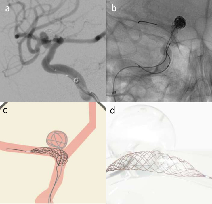
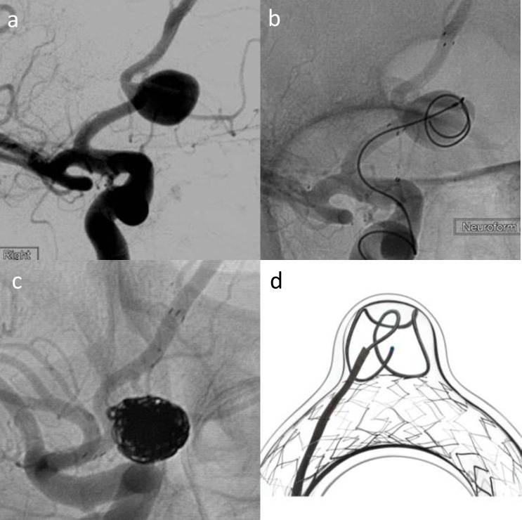
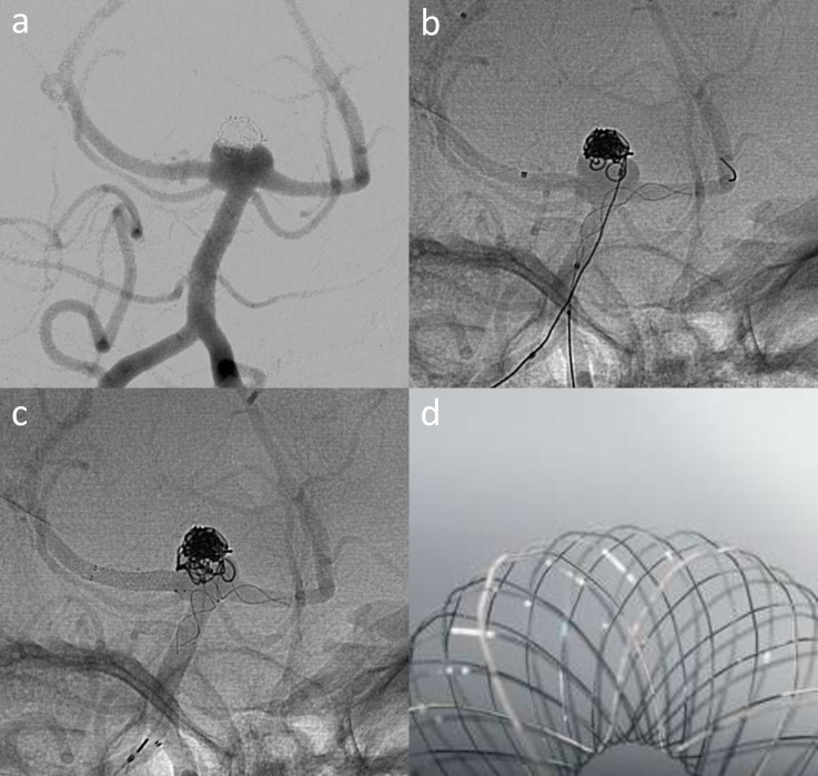
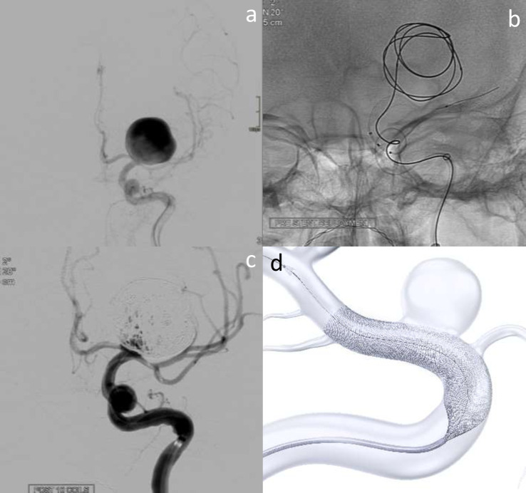
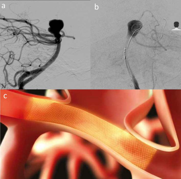
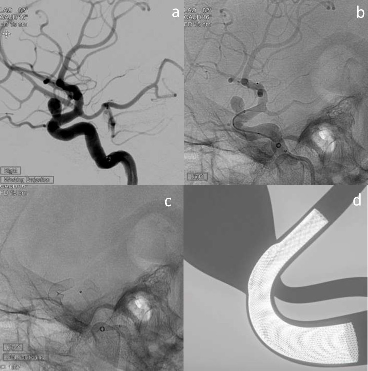
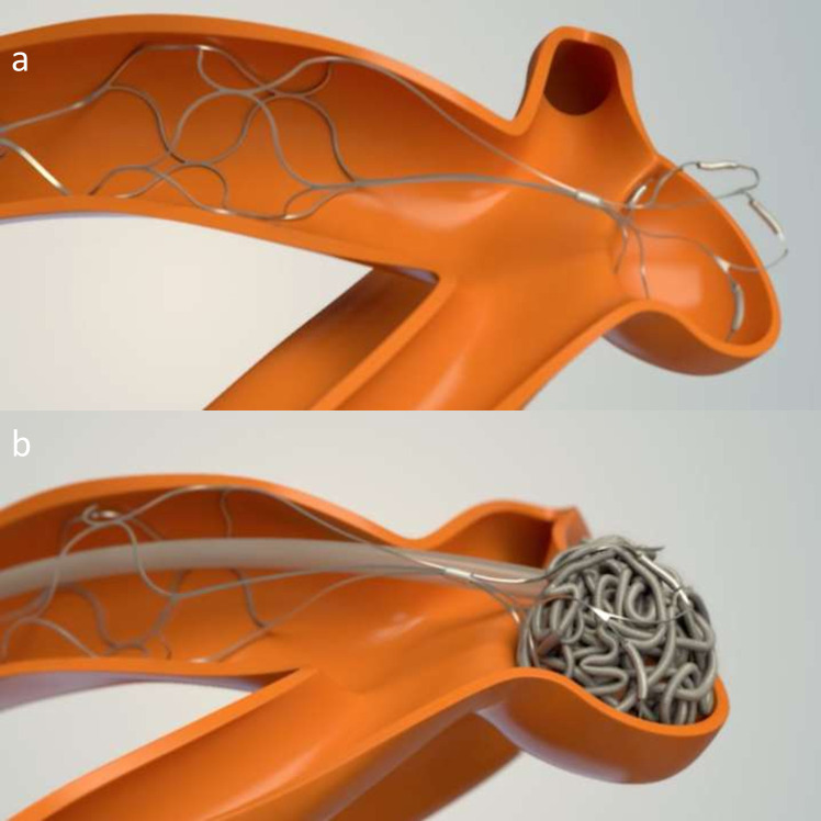
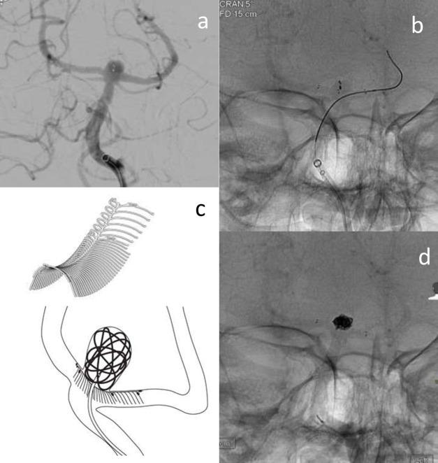
The International Subarachnoid Aneurysm Trial (ISAT) showed superiority for endovascular treatment of ruptured aneurysms and technology has since moved on rapidly. Many approaches and technology now exist for the endovascular management of ruptured and unruptured intracranial aneurysms, which reflects their varied nature - there is no one-size-fits-all technique. We aim to provide an overview of the various classes of device and the major developments over the past decade. Coiling is the oldest of the technology and continues to demonstrate high levels of occlusion and acceptable risks, making it the default treatment choice, particularly in the acutely ruptured aneurysm setting. Advances on coiling include the use of adjuncts such as balloons, stents and fully retrievable temporary neck-bridging devices, which have facilitated the treatment of more complex aneurysms. Flow divertors have also revolutionised complex aneurysm treatment with small added risk in acute aneurysm treatment and seek to remodel the aneurysm-vessel interface without accessing the aneurysm sac. The latest development and most promising avenue appears to be intrasaccular flow disrupting devices like WEB, Contour and Neqstent that provide excellent opportunities to treat wide neck complex aneurysm with minimal mortality and morbidity and good occlusion rates and may in future replace a significant number of stent-assisted coiling too.


Figures

Figure 1.
Figure 1.
Elective re-treatment of a right carotico-ophthalmic aneurysm (a), previously treated with balloon-assisted coiling. Angiography image showing recurrence treated with Comaneci-assisted coiling (b). Illustration demonstrating Comaneci-assisted coiling (c) and a image of the deployed comaneci device (1d) – images provided courtesy of Rapid-Medical.
Figure 2.
Figure 2.
Elective treatment of a wide-neck Acom aneurysm (2a) with Neuroform atlas stent-assisted coiling using a combination of Target 360 Soft, Axium Prime and Kaneka coils (b–d). (b) shows deployment of the first coil with stent across the neck, and 2c shows the final coil mass with stent in situ. (d) is a schematic image illustrating Neuroform atlas-assisted coiling in a model vessel and aneurysm – courtesy of Stryker Neurovascular.
Figure 3.
Figure 3.
LVIS EVO stent shown fully opened. Image provided courtesy of Microvention/Terumo.
Figure 4.
Figure 4.
Elective treatment of a recurrent basilar tip aneurysm (a) that was previously treated with balloon-assisted coiling. T-stenting performed with Baby Leo deployed from the left P1 to the basilar (b) and LVIS EVO across the right P1 (c). A image of the Baby Leo device illustrates the stent cells that support the coil mass – image courtesy of Sela Medical.
Figure 5.
Figure 5.
Surpass Evolve assisted coiling of giant left ICA termination aneurysm. a shows a giant terminal ICA aneurysm on the left. b demonstrates coil deployment into the aneurysm, with Surpass Evolve stent placed in the terminal ICA, but not yet deployed. 5c shows small volume filling after several coils have been deployed. d is a schematic image of the stent during deployment – courtesy of Stryker Neurovascular.
Figure 6.
Figure 6.
Acute admission with lumbar puncture positive SAH. Wide-neck basilar aneurysm (a) treated with Pipeline Embolisation Device with Shield Technology delivered to left P1 (b) and across the aneurysm neck. Schematic illustration (c) of PED shield across an aneurysm neck illustrates the device position – image courtesy of Medtronic.
Figure 7.
Figure 7.
Elective treatment of a right carotico-ophthalmic aneurysm (a) with p64 HPC-coated flow diverter. b shows the initial opening of the flow diverter beyond the aneurysm neck, and c shows the flow diverter in situ and fully deployed. d is a schematic representation of the p64 device in situ across the aneurysm neck – image courtesy of Phenox.
Figure 8.
Figure 8.
pCONus two device mechanism illustrated through schematic images. a shows the device placed in the parent vessel and petals sited within the aneurysm neck and sac. b shows the pCONus two assisted coiling of the aneurysm.
Figure 9.
Figure 9.
E clips deployed into both PCA’s and across the aneurysm neck to facilitate coil placement into a basilar tip aneurysm recurrence (previously treated with WEB). a shows the recurrent basilar tip aneurysm with previous WEB device visible. b shows microcatheter in the left PCA and a deployed eCLIPs device placed across the aneurysm neck. c is a schematic drawing and image of the eCLIPs device showing how it supports coiling of a bifurcation aneurysm – courtesy of Evasc Medical Systems. 9d shows the final coil mass, deployed through the eCLIPs device.
Figure 10.
Figure 10.
WEB placement in an acutely ruptured complex Acom aneurysm with superiorly located rupture point (a). b and c show the deployed WEB device within the aneurysm sac. d is a schematic diagram that shows how the WEB device sits within the aneurysm sac – courtesy of Microvention.
Figure 11.
Figure 11.
Images of the Cerus Contour Neurovascular System. Top row shows the device in the open form. Bottom row shows the device in the deployed device in the “Chalice” shape that conforms to the aneurysm neck. Images courtesy of Cerus.
Figure 12.
Figure 12.
Elective right Acom aneurysm (a) treated with the Contour Neurovascular System (CNS). b shows deployment of the CNS before it is detached. c shows the device in situ across the neck with contrast stasis. d is a schematic figure that shows the device sat in the aneurysm neck – courtesy of Cerus.
Figure 13.
Figure 13.
Previously coiled Acom aneurysm following acute rupture and SAH. Additional unruptued left terminal ICA aneurysm treated with Contour Neurovascular System (CNS) (13a). b shows deployment of the CNS before detachment. c shows the device in-situ across the aneurysm neck. d is a schematic image of the CNS post-deployment with contrast stasis within the the aneurysm – courtesy of Cerus.
Figure 14.
Figure 14.
Acute Grade 1 SAH from left Acom aneurysm (a) treated with Neqstent-assisted coiling. b is a picture of the Neqstent device which sits across the neck and supports coiling and c shows deployment of the Neqstent device (arrowhead) with second microcatheter delivering coils into the aneurysm sac. d is a schematic figure illustrating how the Neqstent supports coiling of bifurcation aneurysms. Images courtesy of Cerus.

Similar articles

Cited by

References

    1. Molyneux AJ, Kerr RSC, Yu L-M, Clarke M, Sneade M, Yarnold JA, et al. . International subarachnoid aneurysm trial (ISAT) of neurosurgical clipping versus endovascular coiling in 2143 patients with ruptured intracranial aneurysms: a randomised comparison of effects on survival, dependency, seizures, rebleeding, subgroups, and aneurysm occlusion. Lancet 2005; 366: 809–17. doi: 10.1016/S0140-6736(05)67214-5 - DOI - PubMed
    1. Ferns SP, Sprengers MES, van Rooij WJ, Rinkel GJE, van Rijn JC, Bipat S, et al. . Coiling of intracranial aneurysms: a systematic review on initial occlusion and reopening and retreatment rates. Stroke 2009; 40: e523-9. doi: 10.1161/STROKEAHA.109.553099 - DOI - PubMed
    1. Pierot L, Wakhloo AK. Endovascular treatment of intracranial aneurysms: current status. Stroke 2013; 44: 2046–54. doi: 10.1161/STROKEAHA.113.000733 - DOI - PubMed
    1. Pierot L, Barbe C, Ferré J-C, Cognard C, Soize S, White P, et al. . Patient and aneurysm factors associated with aneurysm rupture in the population of the ARETA study. J Neuroradiol 2020; 47: 292–300. doi: 10.1016/j.neurad.2019.07.007 - DOI - PubMed
    1. Pierot L, Barbe C, Herbreteau D, Gauvrit J-Y, Januel A-C, Bala F, et al. . Rebleeding and bleeding in the year following intracranial aneurysm coiling: analysis of a large prospective multicenter cohort of 1140 patients-Analysis of recanalization after endovascular treatment of intracranial aneurysm (ARETA) study. J Neurointerv Surg 2020; 12: 1219–25. doi: 10.1136/neurintsurg-2020-015971 - DOI - PubMed