

MRI-directed biopsy for primary detection of prostate cancer in a population of 223 men: MRI In-Bore vs MRI-transrectal ultrasound fusion-targeted techniques
- PMID: 34609900
- PMCID: PMC8978234
- DOI: 10.1259/bjr.20210528
MRI-directed biopsy for primary detection of prostate cancer in a population of 223 men: MRI In-Bore vs MRI-transrectal ultrasound fusion-targeted techniques
Abstract
Objectives: To compare the detection rates of overall prostate cancer (PCa) and clinically significant PCa (csPCa) and the median percentage of cancer per biopsy core between MRI-guided In-bore and MRI-TRUS fusion-targeted biopsy (TBx).
Methods: In this retrospective study, 223 patients who underwent prostate multiparametric MRI (mpMRI) and subsequent MR-directed biopsy were included. For PCa and csPCa detection rate (DR), contingency tables were tested via the Pearson's chi-squared to explore the variance of the outcome distribution. The percentage of cancer per biopsy core was tested with a two-tailed Mann-Withney test.
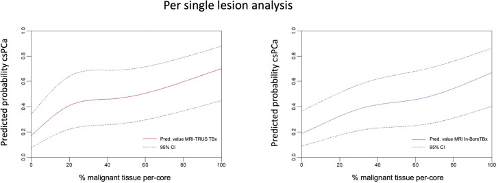
Results: One hundred and seventeen and 106 patients underwent MRI-TRUS fusion or MRI In-bore TBx, respectively. 402 MRI biopsy targets were identified, of which 206 (51.2%) were biopsied with the MRI-TRUS TBx and 196 (48.8%) with the MRI In-bore TBx technique. Per-patient PCa and csPCa detection rates were 140/223 (62.8%) and 97/223 (43.5%), respectively. PCa-DR was 73/117 (62.4%) and 67/106 (63.2%) for MRI-TRUS and MRI In-Bore TBx (p = 0.9), while csPCa detection rate reached 50/117 (42.7%) and 47/106 (44.3%), respectively (p = 0.81). The median per-patient percentage of malignant tissue within biopsy cores was 50% (IQR: 27-65%) for PCa and 60% (IQR: 35-68%) for csPCa, with a statistically significant difference between the techniques.
Conclusion: No statistically significant difference in the detection rate of MRI In-bore and MRI-TRUS fusion TBx was found. MRI In-bore TBx showed higher per-core percentage of malignant cells.
Advances in knowledge: MRI In-bore biopsy might impact risk stratification and patient management considering the higher per-core percentage of malignant cells, especially for patients eligible for active surveillance or focal therapy.
Figures
References
-
- Mottet RCN, van den Bergh N, Brier E, Cornford P, De Santis M, Fanti S. EAU guidelines: prostate cancer. EAU guidelines. 978th-94th-92671st-04-2. ed; 2019.
-
- Faria R, Soares MO, Spackman E, Ahmed HU, Brown LC, Kaplan R, et al. Optimising the diagnosis of prostate cancer in the era of multiparametric magnetic resonance imaging: a cost-effectiveness analysis based on the prostate MR imaging study (PROMIS). Eur Urol 2018; 73: 23–30. doi: 10.1016/j.eururo.2017.08.018 - DOI - PMC - PubMed
-
- Panebianco V, Valerio MC, Giuliani A, Pecoraro M, Ceravolo I, Barchetti G, et al. Clinical utility of multiparametric magnetic resonance imaging as the first-line tool for men with high clinical suspicion of prostate cancer. Eur Urol Oncol 2018; 1: 208–14. doi: 10.1016/j.euo.2018.03.008 - DOI - PubMed
Publication types
MeSH terms
LinkOut - more resources
Full Text Sources
Medical
