Review
. 2022 Jan 1;95(1129):20201241.
doi: 10.1259/bjr.20201241. Epub 2021 Oct 5.

Neuroradiological findings in Alagille syndrome

Affiliations
Review

Neuroradiological findings in Alagille syndrome

Alessandra D'Amico et al. Br J Radiol. .

Abstract

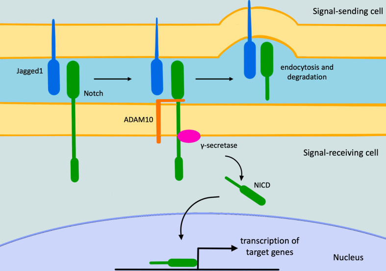
Alagille syndrome (ALGS) is a multisystemic disease caused by mutations in genes of Notch pathway, which regulates embryonic cell differentiation and angiogenesis. Clinically, ALGS is characterized by cholestasis, cardiac defects, characteristic facial features, skeletal and ophthalmologic abnormalities. The aim of this review is to illustrate neuroradiological findings in ALGS, which are less well-known and prevalent, including cerebrovascular anomalies (such as aneurysms, dolichoectasia, Moyamoya syndrome and venous peculiarities), Chiari 1 malformation, craniosynostosis, intracranial hypertension, and vertebral anomalies (namely butterfly vertebra, hemivertebra, and craniocervical junction anomalies). Rarer cerebral midline malformations and temporal bone anomalies have also been described.


Figures

Figure 1.
Figure 1.
The Notch signaling pathway. The pathway starts with Notch/Jagged1 interaction, causing the Adam10 metalloprotease activation. This last cleaves Notch allowing the Notch-Jagged1 complex to be endocytosed by the ligand-expressing cell. Subsequently, the γ-secretase enzyme cleaves the remaining part of Notch releasing its intracellular domain that moves to the nucleus, where it regulates gene expression. NICD, intracellular domain of the Notch protein.
Figure 2.
Figure 2.
Cerebrovascular anomalies in a 9-year-old girl affected by ALGS. Axial 3D-time of flight MRA of the circle of Willis (a, b) depicts a small aneurysm of the cavernous segment of the left internal carotid artery (arrow in a) and narrowing of the petrous segment of the right internal carotid artery (arrowheads in b). ALGS, alagille syndrome; MRA, MR angiography.
Figure 3.
Figure 3.
Cerebral arterial anomalies in a 2-year-old boy with ALGS. Coronal 3D-time of flight MRA of the circle of Willis shows stenotic narrowing of the carotid siphons (arrows). ALGS, alagille syndrome; MRA, MR angiography.
Figure 4.
Figure 4.
Cerebral venous anomalies in a 15-year-old boy with ALGS. Coronal 3D-phase contrast MRA shows diffuse tortuosity of cerebral veins compared to a child of comparable age without ALGS (b). ALGS, alagille syndrome; MRA, MR angiography.
Figure 5.
Figure 5.
Chiari 1 in a 17-year-old boy with ALGS. Sagittal (a) and axial (b) turbo spin echo T2 weighted images depicting low lying cerebellar tonsils (white arrow in A) and reduced volume of the posterior fossa (b). Clivus and cervical vertebral bodies are hypoplastic (black arrows in A). Note also scaphocephaly and frontal bossing. ALGS, alagille syndrome
Figure 6.
Figure 6.
IIH in a 17-year-old boy with ALGS suffering from chronic headache. Axial turbo spin echo T2 weighted image (a) and coronal 3D-phase contrast MRA (b) depict dilation of the optic nerve sheaths (black arrows in A) and a signal gap in the right transverse sinus (white arrows in b) due to stenosis, respectively. Also note flattening of the posterior globes contour (a). ALGS, alagille syndrome; IIH, idiopathic intracranial hypertension; MRA, MR angiography.
Figure 7.
Figure 7.
Basicranium anomalies in a 14-year-old girl with ALGS. Sagittal (a) and axial (b) turbo spin echo T2 weighted images show foveola pharyngica recess (white arrows in A and B) and clival hypoplasia (black arrow in a). ALGS, alagille syndrome
Figure 8.
Figure 8.
Cerebral midline anomalies in a 15-year-old-girl with ALGS. Coronal turbo spin echo T2 weighted image shows isolated agenesis of the septum pellucidum (a). Cerebral midline and hippocampal anomalies in a 14-year-old girl with ALGS. Coronal (b) and sagittal (c) turbo spin echo T2 weighted images demonstrate bilateral incomplete hippocampal inversion (black arrows in b) and mild hypoplasia of the splenium and the isthmus of the corpus callosum (black arrowheads in c). ALGS, alagille syndrome
Figure 9.
Figure 9.
Spine malformations in a 35-year-old male with ALGS. Coronal turbo spin echo T2 weighted image depicts severe vertebral dysmorphism, consisting of C5 and C6 fusion (segmentation anomaly) and typical median split of the vertebral body (butterfly vertebra) (white arrows). ALGS, alagille syndrome
Figure 10.
Figure 10.
Spine malformation in a 10-year-old boy with ALGS. Coronal turbo spin echo T2 weighted images (a–c) depict a left thoracic hemivertebra (arrows in a–c). ALGS, alagille syndrome

Similar articles

Cited by

References

    1. Danks DM, Campbell PE, Jack I, Rogers J, Smith AL. Studies of the aetiology of neonatal hepatitis and biliary atresia. Arch Dis Child 1977; 52: 360–7. doi: 10.1136/adc.52.5.360 - DOI - PMC - PubMed
    1. Saleh M, Kamath BM, Chitayat D. Alagille syndrome: clinical perspectives. Appl Clin Genet 2016; 9: 75–82. doi: 10.2147/TACG.S86420 - DOI - PMC - PubMed
    1. D'Souza B, Miyamoto A, Weinmaster G. The many facets of Notch ligands. Oncogene 2008; 27: 5148–67. doi: 10.1038/onc.2008.229 - DOI - PMC - PubMed
    1. Mitchell E, Gilbert M, Loomes KM. Alagille syndrome. Clin Liver Dis 2018; 22: 625–41. doi: 10.1016/j.cld.2018.06.001 - DOI - PubMed
    1. Carpenter CD, Linscott LL, Leach JL, Vadivelu S, Abruzzo T. Spectrum of cerebral arterial and venous abnormalities in Alagille syndrome. Pediatr Radiol 2018; 48: 602–8. doi: 10.1007/s00247-017-4043-2 - DOI - PubMed