. 2021 Nov 15:712:109051.
doi: 10.1016/j.abb.2021.109051. Epub 2021 Oct 2.

Rapid preparation of nanodiscs for biophysical studies

Affiliations

Rapid preparation of nanodiscs for biophysical studies

Jeffrey A Julien et al. Arch Biochem Biophys. .

Erratum in

Abstract

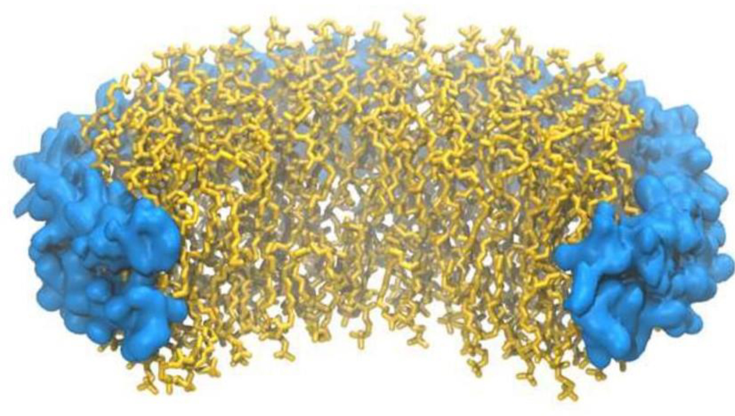
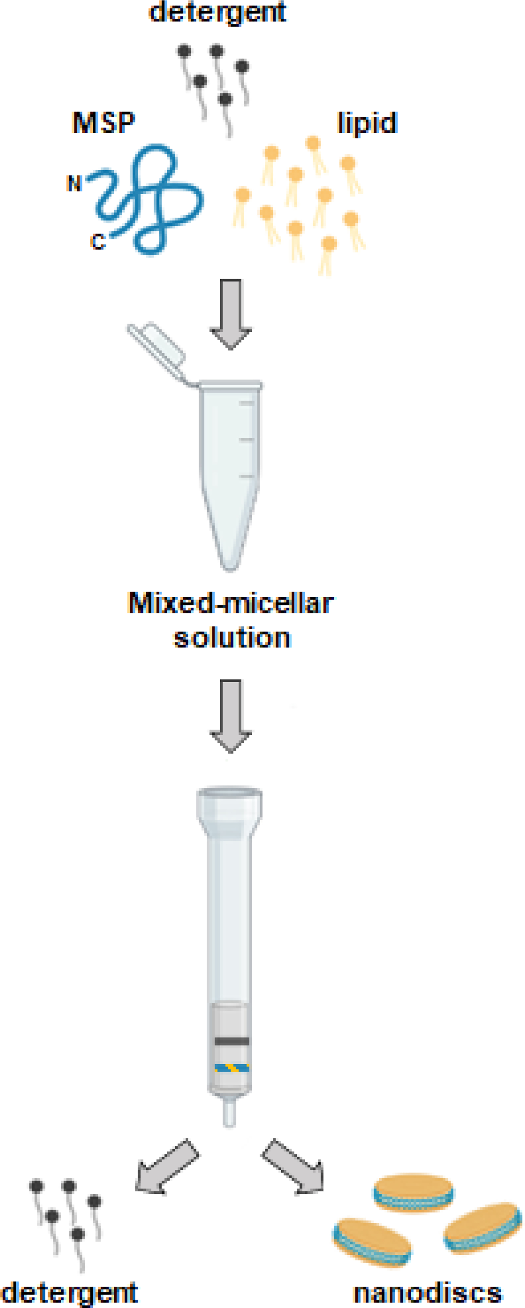
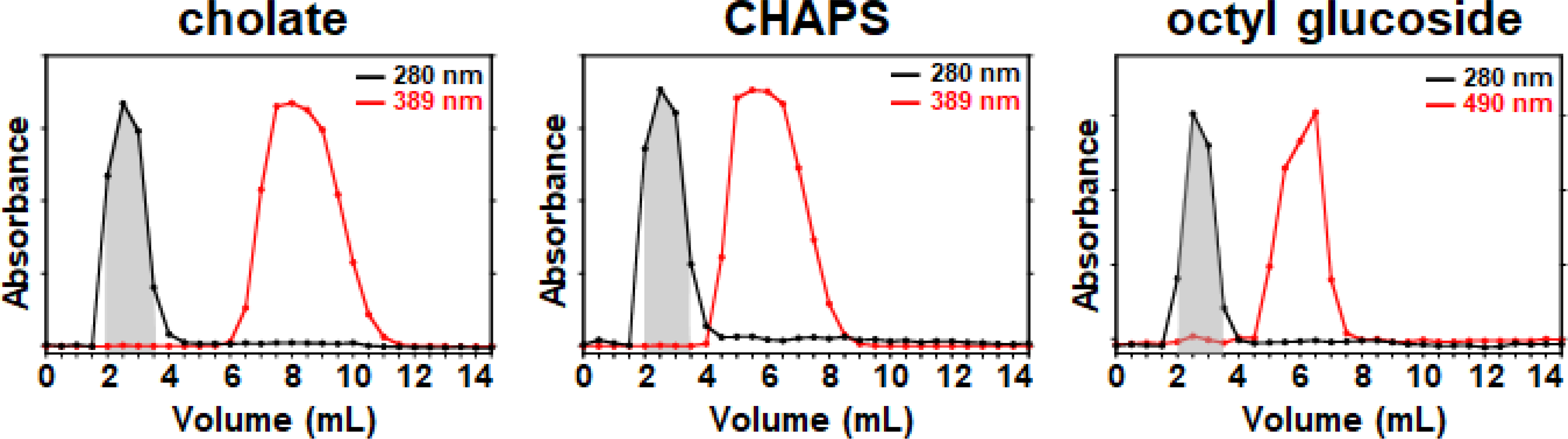
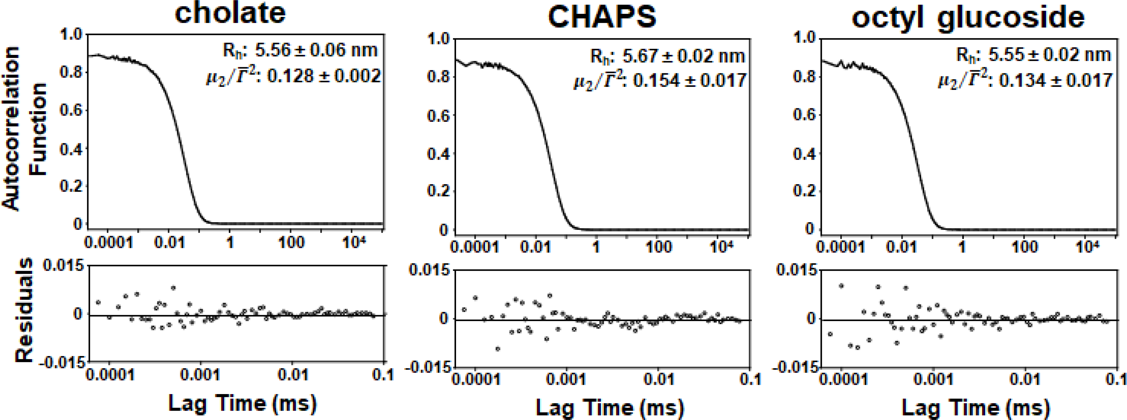
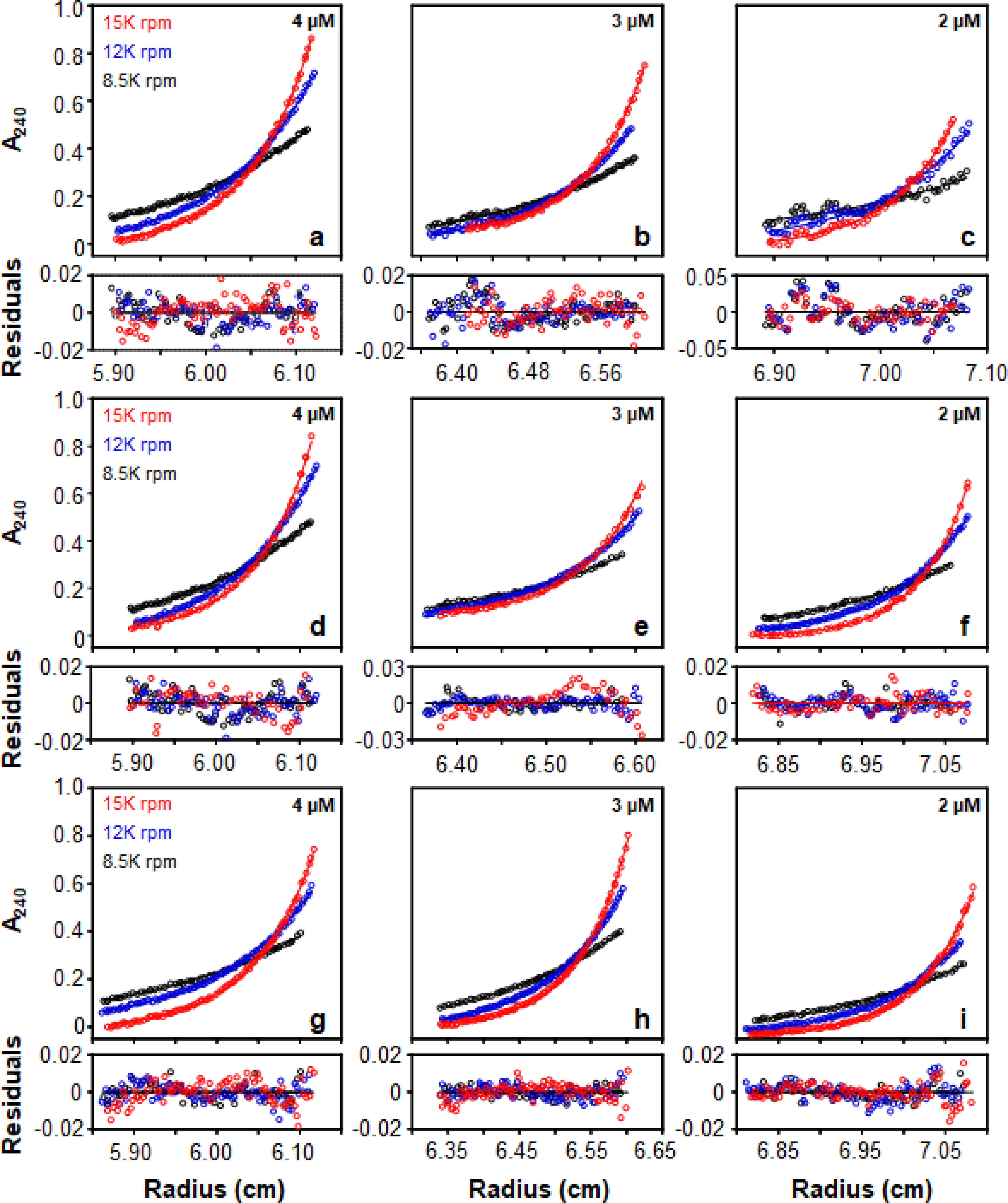
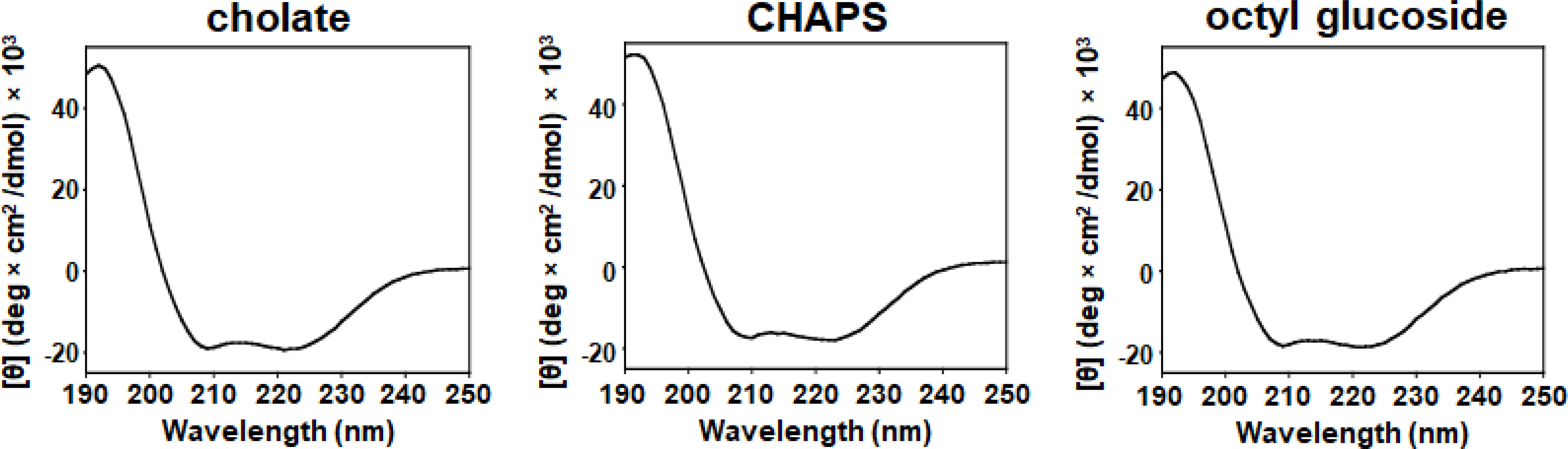
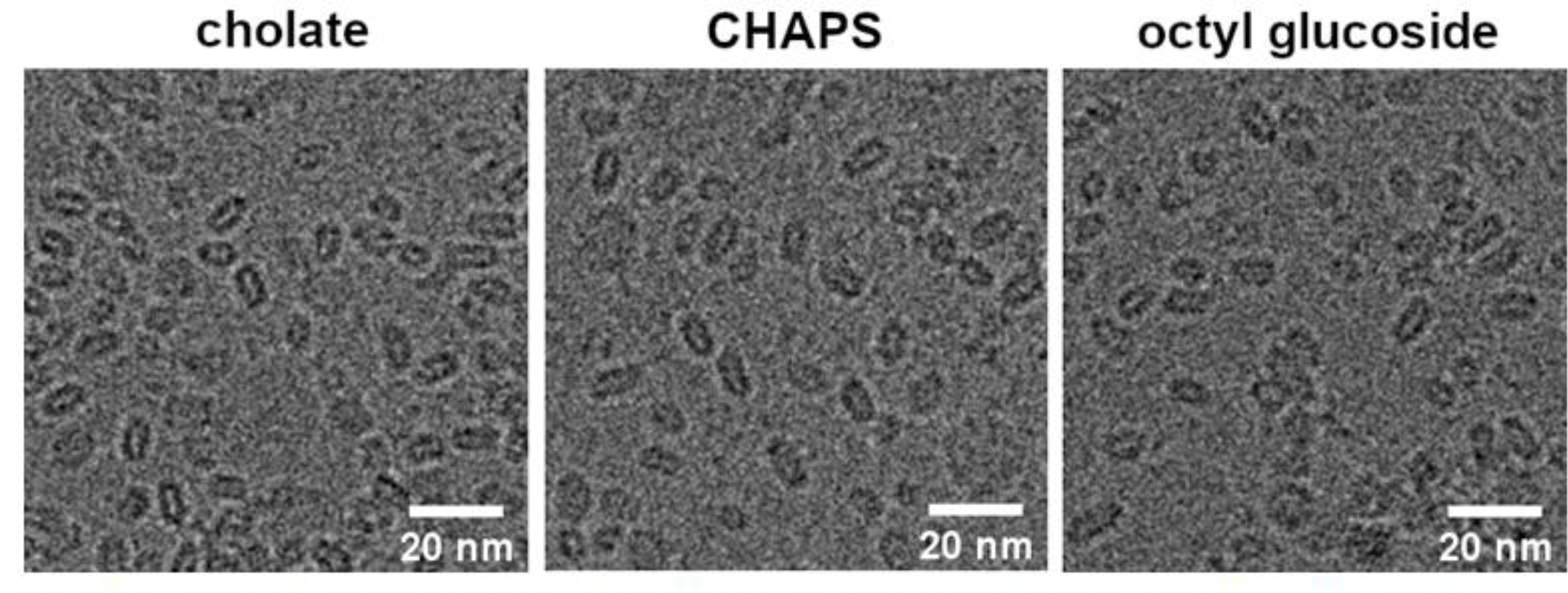
Nanodiscs, which are disc-shaped entities that contain a central lipid bilayer encased by an annulus of amphipathic helices, have emerged as a leading native-like membrane mimic. The current approach for the formation of nanodiscs involves the creation of a mixed-micellar solution containing membrane scaffold protein, lipid, and detergent followed by a time consuming process (3-12 h) of dialysis and/or incubation with sorptive beads to remove the detergent molecules from the sample. In contrast, the methodology described herein provides a facile and rapid procedure for the preparation of nanodiscs in a matter of minutes (<15 min) using Sephadex® G-25 resin to remove the detergent from the sample. A panoply of biophysical techniques including analytical ultracentrifugation, dynamic light scattering, gel filtration chromatography, circular dichroism spectroscopy, and cryogenic electron microscopy were employed to unequivocally confirm that aggregates formed by this method are indeed nanodiscs. We believe that this method will be attractive for time-sensitive and high-throughput experiments.

Keywords: Analytical ultracentrifugation; Circular dichroism spectroscopy; Cryogenic electron microscopy; Dynamic light scattering; Nanodiscs; Reconstitution.


Conflict of interest statement

Conflicts of interests.

The authors declare that there are no competing conflicts of interest.

Figures

Figure 1.
Figure 1.
Cartoon image of a nanodisc. Membrane scaffolding protein is shown in blue. Dimyristoylphosphatidylcholine lipid is shown in yellow.
Figure 2.
Figure 2.
Pictorial representation of hypothetical nanodisc formation using Sephadex® G-25 resin.
Figure 3.
Figure 3.
Elution profiles of mixed micellar solutions passed over Sephadex® G-25 resin. Spherical data points represent elution fractions. Red traces indicate the elution profile of detergent. Absorbances are normalized for facile viewing. 10 mM KH2PO4 pH 7.4, 50 mM Na2SO4 was used to equilibrate the column and collect fractions.
Figure 4.
Figure 4.
Elution profiles of particles on a HiLoad™ 16/60 Superdex™ 200 column. vo is void volume and ve is elution volume. 10 mM KH2PO4 pH 7.4, 50 mM Na2SO4 was used to equilibrate the column and collect fractions.
Figure 5.
Figure 5.
DLS autocorrelation functions, Rh, and μ2/Γ¯2 values of the two highest intensity fractions from the Superdex™ 200 elution profile of particles produced on the Sephadex® G-25 column using 1× phosphate-buffered saline (PBS). Data was collected for 5 mins at 25° C using values of viscosity and refractive index of 1.0200 cP and 1.335 respectively for PBS.[35]
Figure 6.
Figure 6.
Sedimentation equilibrium profiles of particles produced on the Sephadex® G-25 column using sodium cholate (a-c), CHAPS (d-f), and octyl glucoside (g-i) in 10 mM KH2PO4 pH 7.4, 50 mM Na2SO4 at 25°C. Residuals are displayed underneath each equilibrium profile.
Figure 7.
Figure 7.
Circular dichroism spectra of particles in 10 mM KH2PO4 pH 7.4, 50 mM Na2SO4 at 25°C. [Θ] is mean residue ellipticity.
Figure 8.
Figure 8.
Cryogenic electron microscopy images of particles produced using Sephadex® G-25 resin with 10 mM KH2PO4 pH 7.4, 50 mM Na2SO4.
Figure 9.
Figure 9.
Particle reconstruction images of particles produced using Sephadex® G-25 resin with 10 mM KH2PO4 pH 7.4, 50 mM Na2SO4.

References

    1. Denisov IG, Sligar SG, Nanodiscs in Membrane Biochemistry and Biophysics, Chem. Rev. 117 (2017) 4669–4713. 10.1021/acs.chemrev.6b00690. - DOI - PMC - PubMed
    1. Boldog T, Li M, Hazelbauer GL, [14] - Using Nanodiscs to Create Water-Soluble Transmembrane Chemoreceptors Inserted in Lipid Bilayers, in: Simon MI, Crane BR, Crane A (Eds.), Meth. Enzymol, Academic Press, 2007: pp. 317–335. 10.1016/S0076-6879(07)23014-9. - DOI - PubMed
    1. Yokogawa M, Fukuda M, Osawa M, Nanodiscs for Structural Biology in a Membranous Environment, Chem. Pharm. Bull. (Tokyo). 67 (2019) 321–326. 10.1248/cpb.c18-00941. - DOI - PubMed
    1. Sun C, Gennis RB, Single-particle cryo-EM studies of transmembrane proteins in SMA copolymer nanodiscs, Chem. Phys. Lipids. 221 (2019) 114–119. 10.1016/j.chemphyslip.2019.03.007. - DOI - PMC - PubMed
    1. Raschle T, Hiller S, Yu T-Y, Rice AJ, Walz T, Wagner G, Structural and Functional Characterization of the Integral Membrane Protein VDAC-1 in Lipid Bilayer Nanodiscs, J. Am. Chem. Soc. 131 (2009) 17777–17779. 10.1021/ja907918r. - DOI - PMC - PubMed

Publication types