. 2021 Nov;40(47):6513-6526.
doi: 10.1038/s41388-021-01872-9. Epub 2021 Oct 5.

RING-finger protein 6 promotes colorectal tumorigenesis by transcriptionally activating SF3B2

Affiliations

RING-finger protein 6 promotes colorectal tumorigenesis by transcriptionally activating SF3B2

Hui Xu et al. Oncogene. 2021 Nov.

Abstract

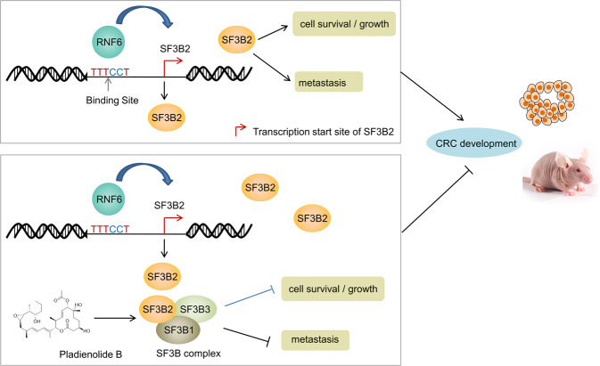
RNF6 is a RING finger protein with oncogenic potential. In this study, we established colon-specific RNF6 transgenic (tg) mice, and demonstrated that RNF6 overexpression accelerated colorectal carcinogenesis compared to wild-type littermates in a chemically induced colorectal cancer (CRC) model. To understand whether transcriptional activity of RNF6 underlies its oncogenic effect, we performed integrated chromatin immunoprecipitation (ChIP)-sequencing and RNA-sequencing analysis to identify splicing factor 3b subunit 2 (SF3B2) as a potential downstream target of RNF6. RNF6 binds to the SF3B2 promoter and the overexpression of RNF6 activates SF3B2 expression in CRC cells, primary CRC organoids, and RNF6 tg mice. SF3B2 knockout abrogated the tumor promoting effect of RNF6 overexpression, whereas the reexpression of SF3B2 recused cell growth and migration/invasion in RNF6 knockout cells, indicating that SF3B2 is a functional downstream target of RNF6 in CRC. Targeting of RNF6-SF3B2 axis with SF3B2 inhibitor with pladienolide B suppressed the growth of CRC cells with RNF6 overexpression in vitro and in vivo. Moreover, the combination of 5-fluorouracil (5-FU) plus pladienolide B exerted synergistic effects in CRC with high RNF6 expression, leading to tumor regression in xenograft models. These findings indicate that tumor promoting effect of RNF6 is achieved mainly via transcriptional upregulation of SF3B2, and that RNF6-SF3B2 axis is a promising target for CRC therapy.


Conflict of interest statement

The authors declare no competing interests.

Figures

Fig. 1
Fig. 1. Colon-specific tg RNF6 expression accelerates AOM-induced CRC in mice.
A Scheme for the generation of mice with colon-specific RNF6 transgenic (tg) expression. B Experimental design for AOM-induced CRC in mice. The numbers of wild-type and RNF6 tg mice are 13 and 11, respectively. C Western blot and RT-qPCR confirmed overexpression of RNF6 in the colon of RNF6 tg mice. GAPDH was used as a loading control. D Representative dissection micrographs of colon tumors from AOM-induced CRC in mice. E H&E staining of wild-type and RNF6 tg mouse colons. Colon-specific RNF6 overexpression increased tumor incidence and extent of dysplasia. F CRC tumor incidence, tumor burden, and percentage of mice displaying the highest extent of dysplasia in wild-type and RNF6 tg mice. LGD low-grade dysplasia, HGD high-grade dysplasia. G Ki-67 staining of cell proliferation in wild-type and RNF6 tg mice. Colon-specific RNF6 overexpression increased cell proliferation. H TUNEL analysis of apoptosis in wild-type and RNF6 tg mice. Colon-specific RNF6 overexpression reduced apoptosis. Data were expressed as mean ± SD.
Fig. 2
Fig. 2. SF3B2 is a transcriptional target of RNF6 via the integrated analysis of ChIP-sequencing and RNA-sequencing.
A Confocal microscope image of RNF6 localization by immunofluorescence (scale bar 10 μm). B Candidate genes were selected based on: (1) ChIP-seq RNF6 binding peaks with 5000 bp of TSS and (2) differential expression (>1.5-fold) in RNA-seq dataset. C mRNA expression of candidate genes upon RNF6 overexpression in HCT116 and HT29 by qPCR (n = 3, performed in triplicate). Only SF3B2 mRNA was consistently increased in HCT116 and HT29 cells overexpressing RNF6. D Expression of SF3B2 upon RNF6 knockout in DLD1 and SW480 cells by Western blot. GAPDH was used as a loading control. E Schematic figure summarizing RNF6 ChIP-PCR primer sets in the SF3B2 promoter region (upper). ChIP assay was performed in SW480 cells to confirm RNF6 binding to the SF3B2 promoter (lower left). ChIP-qPCR validation of RNF6 enrichment in SF3B2 promoter (lower right). F EMSA performed with biotin-labeled SF3B2 probes and SW480 nuclear extracts. Specific protein–DNA complexes are indicated by arrows. NSB nonspecific binding. Competitive EMSA result demonstrated that nuclear extracts from SW480 cells induced bands shift for SF3B2. In supershift assay, low or high dose of RNF6 antibody and IgG were added to reaction mixture after the reaction of nuclear extract and SF3B2 probes. Supershifts by anti-RNF6 antibody are indicated with the red arrows. G RNF6-induced SF3B2 promoter reporter activity, as determined by dual-luciferase reporter assay (n = 3, performed in triplicate). Data were expressed as mean ± SD. *P < 0.05, **P < 0.01, ***P < 0.001.
Fig. 3
Fig. 3. RNF6 upregulates SF3B2, a novel oncogenic factor in CRC.
A SF3B2 protein and mRNA expression in colon tissues from wild-type and RNF6 tg mice was determined by Western blot and RT-qPCR, respectively. Ectopic expression of RNF6 increased SF3B2 protein and mRNA in the RNF6 tg mice colon. GAPDH was used as a loading control. B RNF6 mRNA positively correlated with SF3B2 mRNA in our cohort (n = 151) and TCGA dataset (n = 623). C mRNA expression of SF3B2 in TCGA cohort. SF3B2 mRNA levels were significantly higher in CRC tumor tissues (n = 623) compared with normal tissue (n = 51) from TCGA cohort. D IHC staining of colon tissues for SF3B2 and quantification of IHC staining in normal colon tissue and paired CRC tumors (n = 16). E SF3B2 protein expression in 205 CRC tissues from our cohort was assessed by IHC staining (left). Kaplan–Meier survival curve of overall survival according to SF3B2 nuclear expression in CRC tissues (right). F Kaplan–Meier survival curve of overall survival according to SF3B2 mRNA expression in TCGA cohort. CRC patients with high SF3B2 protein expression had significantly shorter survival. G Cell viability was decreased after SF3B2 knockout with sgRNA in DLD1 and SW480 cells (n = 3, performed in triplicate). H SF3B2 knockout suppressed colony formation in DLD1 and SW480 cells (n = 3, performed in triplicate). I Effect of SF3B2 knockout on cell migration by transwell assay in DLD1 and SW480 cells (n = 3, performed in triplicate). J Effect of SF3B2 knockout on cell invasion by Matrigel-coated transwell assay in DLD1 and SW480 cells (n = 3, performed in triplicate). Data were expressed as mean ± SD. **P < 0.01. ***P < 0.001.
Fig. 4
Fig. 4. Oncogenic effect of RNF6 in CRC is dependent on SF3B2.
A SF3B2 knockout and wild-type CRC cells (HCT116 and HT29) were transfected with empty vector or RNF6 overexpression plasmid. RNF6 and SF3B2 protein expression were determined by Western blot. GAPDH was used as a loading control. B MTT, C colony formation. D, E Transwell migration and invasion assays (n = 3, performed in triplicate) were used to assess tumorigenic functions. These functional assays collectively demonstrated that SF3B2 knockout abolished the effect of RNF6 overexpression on cell proliferation, colony formation, and cell migration and invasion. Data were expressed as mean ± SD. **P < 0.01. ***P < 0.001.
Fig. 5
Fig. 5. SF3b complex inhibitor pladienolide B suppresses the growth of RNF6-overexpressing tumors in vitro and in vivo.
A Pladienolide B suppressed SF3B2 protein expression in RNF6-overexpressing HCT116 and HT29 cells as determined by Western blot (left). GAPDH was used as a loading control. Plad-B (1 nM) had no effect on SF3B2 mRNA expression (right). B Relative cell viability of vector or RNF6-overexpressing HCT116 and HT29 cells administrated pladienolide B (1 nM) or vehicle (DMSO) (n = 3, performed in triplicate). Viability of cells with RNF6 overexpression was reduced compared to vector controls. C Two human CRC organoids (PDO74 and PDO828) were successfully transfected with empty vector or RNF6 plasmid. GAPDH was used as a loading control. D, E Empty vector or RNF6-overexpressing CRC organoids were photographed after treatment with pladienolide B or vehicle (DMSO), and then quantified by cell viability assay (scale bar 200 μm) (n = 3, performed in triplicate). The cell viability was normalized to untreated control groups. F Pladienolide B (1 or 2 nM) treatment in RNF6-overexpressing PDO74 or PDO828 reduced SF3B2 protein expression, but had no effect on RNF6 protein. GAPDH was used as a loading control. G Tumor volume in nude mice bearing HCT116 cells with empty vector or RNF6 overexpression (n = 5). H Tumor volume and tumor weight in nude mice bearing HCT116 cells with empty vector or RNF6 overexpression were treated with pladienolide B at 3 mg/kg (n = 5). Pladienolide B treatment resulted in greater regression in tumors with stable RNF6 overexpression compared with tumors with empty vector. Data were expressed as mean ± SD. **P < 0.01. ***P < 0.001.
Fig. 6
Fig. 6. Pladienolide B demonstrates synergistic effects with 5-FU in CRC with high RNF6 expression.
A Dose-response curve of 5-FU in empty vector or RNF6-expressing HCT116 and HT29 cells. Cells were treated with 5-FU for 72 h. IC50 values were significantly increased in cells with RNF6 overexpression compared with the control group. B Isobologram analysis of combined effect of 5-FU and pladienolide B (left). RNF6-overexpressing HCT116 and HT29 cells were treated with 5-FU, pladienolide B, or their combination as indicated to determine the interaction of drugs. IC50 and confidence interval (CI) values were shown in the right table. CI values were calculated for quantitative definition of synergism (CI < 1), additive effect (CI = 1) and antagonism (CI > 1). C, D RNF6-overexpressing CRC organoids (PDO74 and PDO828) were photographed after treatment with 5-FU, pladienolide B, or their combination (scale bar 200 μm). Cell viability was normalized to the untreated control groups (n = 3, performed in triplicate). The results showed that proliferation of PDO74 and PDO828 was significantly decreased by combination treatment. E, F Pladienolide B synergized with 5-FU to suppress the growth of HCT116-RNF6 xenografts in nude mice (n = 5). Tumor volume and tumor weight in nude mice bearing HCT116-RNF6 xenografts and given control, 5-FU, pladienolide B, or their combination are shown in (E) and (F). Data were expressed as mean ± SD.
Fig. 7
Fig. 7. Proposed mechanistic scheme of RNF6-SF3B2 axis in CRC.
Our study showed that RNF6 transcriptionally upregulates SF3B2 expression via direct binding to the SF3B2 promoter. Downstream of RNF6, SF3B2 functions as an oncogene that mediates colorectal tumorigenesis. Pladienolide B, an inhibitor of the SF3b complex, is strongly effective in inhibition of tumor growth in CRC with RNF6 overexpression.

References

    1. Siegel RL, Miller KD. Cancer statistics. CA Cancer J Clin. 2020;70:7–30. doi: 10.3322/caac.21590. - DOI - PubMed
    1. Siegel RL, Miller KD. Colorectal cancer statistics. CA Cancer J Clin. 2020;70:145–64. doi: 10.3322/caac.21601. - DOI - PubMed
    1. Watson IR, Takahashi K, Futreal PA, Chin L. Emerging patterns of somatic mutations in cancer. Nat Rev Genet. 2013;14:703–18. doi: 10.1038/nrg3539. - DOI - PMC - PubMed
    1. Fearon ER. Molecular genetics of colorectal cancer. Ann Rev Pathol. 2011;6:479–507. doi: 10.1146/annurev-pathol-011110-130235. - DOI - PubMed
    1. Shen L, Toyota M, Kondo Y, Lin E, Zhang L, Guo Y, et al. Integrated genetic and epigenetic analysis identifies three different subclasses of colon cancer. Proc Natl Acad Sci USA. 2007;104:18654–9. doi: 10.1073/pnas.0704652104. - DOI - PMC - PubMed

Publication types

MeSH terms