

Paraspinal muscle morphology and composition in adolescent idiopathic scoliosis: A histological analysis
- PMID: 34611591
- PMCID: PMC8479518
- DOI: 10.1002/jsp2.1169
Paraspinal muscle morphology and composition in adolescent idiopathic scoliosis: A histological analysis
Abstract
Background: Adolescent idiopathic scoliosis (AIS) is a condition resulting in spinal deformity and tissue adaptation of the paraspinal muscles. Although prior studies have demonstrated asymmetries in fiber type and other energetic features of muscle on the concave side of the curve, muscle morphology, architecture, and composition have not been evaluated. Therefore, the purpose of this study was to compare differences in paraspinal muscle microarchitecture and composition between concave and convex sides of a scoliotic curve in individuals with AIS.
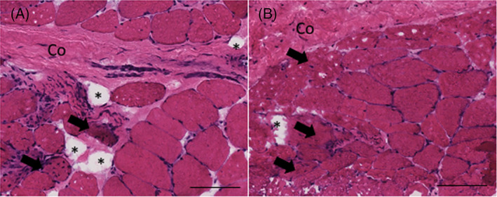
Methods: Paraspinal muscle biopsies were obtained at the apex of the scoliotic curve in 29 individuals with AIS undergoing surgical deformity correction. Histological assays were performed to quantify fiber size, evidence of muscle degeneration and regeneration, and tissue composition (proportion of muscle, collagen, and fat). Differences between contralateral muscle samples were compared using two-tailed paired Student's t tests, and relationships between clinical characteristics (age and curve severity) and muscle characteristics were investigated using Pearson correlations.
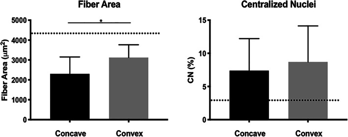
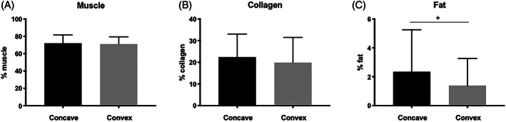
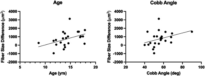
Results: Muscle fibers were significantly larger on the convex side of the curve apex (P = .001), but were lower than literature-based norms for healthy paraspinal muscle. There were no differences in amount of degeneration/regeneration (P = .490) or the proportion of muscle and collagen (P > .350) between the concave and convex sides, but high levels of collagen were observed. There was a trend toward higher fat content on the concave side (P = .074). Larger fiber size asymmetries were associated with greater age (r = .43, P = .046), and trended toward an association with greater curve severity (r = .40, P = .069).
Conclusions: This study demonstrates that although muscle fibers are larger on the convex side of the scoliotic curve in AIS, muscles on both sides are atrophic compared to non-scoliotic individuals, and demonstrate levels of collagen similar to severe degenerative spinal pathologies. These findings provide insight into biological maladaptations occurring in paraspinal muscle in the presence of AIS.
Keywords: adolescent idiopathic scoliosis; deformity; muscle; spine.
© 2021 The Authors. JOR Spine published by Wiley Periodicals LLC on behalf of Orthopaedic Research Society.
Conflict of interest statement
B. S. receives consultation fees from San Diego Spine Foundation and research support from the National Institutes of Health and the Foundation of Physical Therapy Research. P. O. N. receives research support from; DePuy Synthes Spine, Setting Scoliosis Straight Foundation, and MAZOR Surgical Technologies; royalties from DePuy Synthes, Stryker K2M, and Theime Publishing; stocks in ElectroCore; and is a board member for the Scoliosis Research Society, Setting Scoliosis Straight Foundation, Harms Study group, and International Pediatric Orthopedic Think Tank. S. R. W. receives research support from the National Institutes of Health, and is a board member for the San Diego Spine Foundation.
Figures
References
-
- Burwell RG. Aetiology of idiopathic scoliosis: current concepts. Pediatr Rehabil. 2003;6(3–4):137‐170. - PubMed
-
- Cheng JC, Castelein RM, Chu WC, et al. Adolescent idiopathic scoliosis. Nat Rev Dis Primers. 2015;1:15030. - PubMed
-
- Bylund P, Jansson E, Dahlberg E, Eriksson E. Muscle fiber types in thoracic erector spinae muscles. Fiber types in idiopathic and other forms of scoliosis. Clin Orthop Relat Res. 1987;214:222‐228. - PubMed
-
- Ford DM, Bagnall KM, McFadden KD, Greenhill BJ, Raso VJ. Paraspinal muscle imbalance in adolescent idiopathic scoliosis. Spine. 1984;9(4):373‐376. - PubMed
-
- Gonyea WJ, Moore‐Woodard C, Moseley B, Hollmann M, Wenger D. An evaluation of muscle pathology in idiopathic scoliosis. J Pediatr Orthop. 1985;5(3):323‐329. - PubMed
LinkOut - more resources
Full Text Sources
Miscellaneous
