[Preprint]. 2021 Sep 24:2021.09.23.21263047.
doi: 10.1101/2021.09.23.21263047.

Anti-PF4 VITT antibodies are oligoclonal and variably inhibited by heparin

Affiliations

Anti-PF4 VITT antibodies are oligoclonal and variably inhibited by heparin

B Singh et al. medRxiv. .

Update in

Abstract

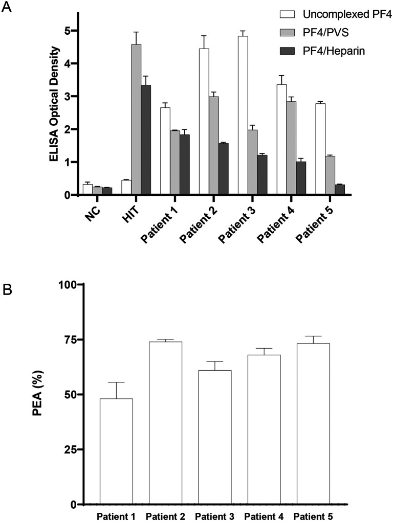
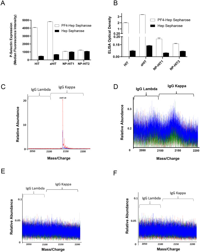
Background: COVID-19 vaccines have been associated with a rare thrombotic and thrombocytopenic reaction, Vaccine-induced immune thrombotic thrombocytopenia (VITT) characterized by platelet-activating anti-PF4 antibodies. This study sought to assess clonality of VITT antibodies and evaluate their characteristics in antigen-based and functional platelet studies.

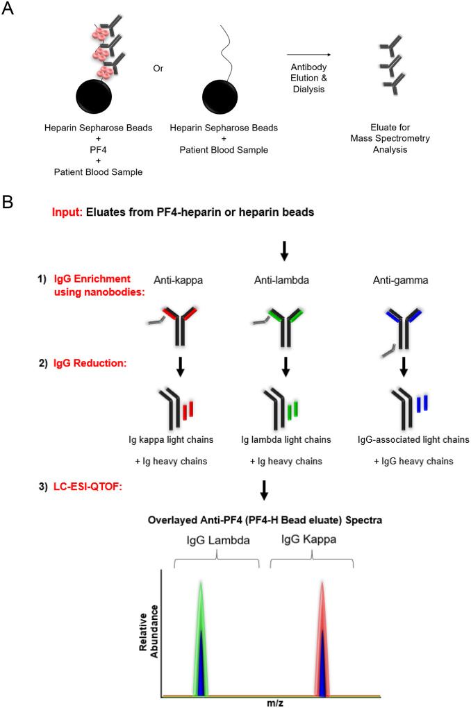
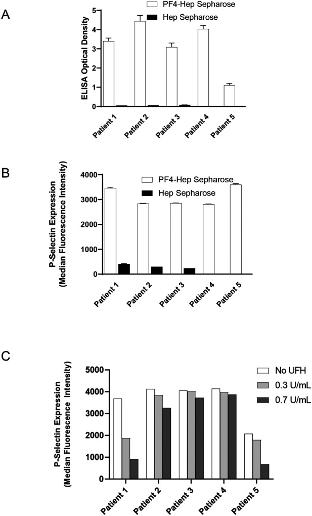
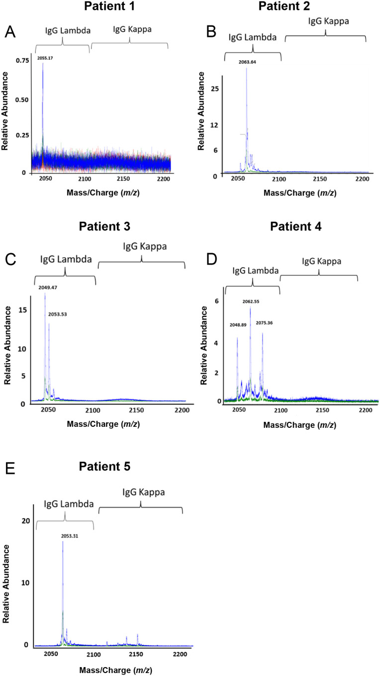
Methods: Anti-PF4 antibodies were isolated from five patients with VITT secondary to ChAdOx1 nCoV-19 (n=1) or Ad26.COV2.S (n=4) vaccination. For comparative studies with heparin-induced thrombocytopenia (HIT), anti-PF4 antibodies were isolated from one patient with spontaneous HIT, another with "classical" HIT, and two patients with non-pathogenic (non-platelet activating) anti-PF4 antibodies. Isolated antibodies were subject to ELISA and functional testing, and mass spectrometric evaluation for clonality determination.

Results: All five VITT patients had oligoclonal anti-PF4 antibodies (3 monoclonal, one bi- and one tri-clonal antibodies), while HIT anti-PF4 antibodies were polyclonal. Notably, like VITT antibodies, anti-PF4 antibodies from a spontaneous HIT patient were monoclonal. The techniques employed did not detect non-pathogenic anti-PF4 antibodies. The ChAdOx1 nCoV-19-associated VITT patient made an excellent recovery with heparin treatment. In vitro studies demonstrated strong inhibition of VITT antibody-induced platelet activation with therapeutic concentrations of heparin in this and one Ad26.COV2.S-associated VITT patient. Oligoclonal VITT antibodies with persistent platelet-activating potential were detected at 6 and 10 weeks after acute presentation in two patients tested. Two of the 5 VITT patients had recurrence of thrombocytopenia and one patient had focal seizures several weeks after acute presentation.

Conclusion: Oligoclonal anti-PF4 antibodies mediate VITT. Heparin use in VITT needs to be further studied.


Conflict of interest statement

Conflict-of-interest disclosure:

AP reports pending/issued patents (Mayo Clinic, Retham Technologies and Versiti BloodCenter of Wisconsin), equity ownership in Retham Technologies, and serving on the advisory board of Veralox Therapeutics. DM reports pending/issued patents (Mayo Clinic). The remaining authors declare no competing financial interests.

Figures

Figure 1.
Figure 1.. VITT antibodies recognize uncomplexed PF4 and activate PF4-treated platelets.
(A) Antibody binding to PF4 alone (“uncomplexed”; white), PF4/Polyvinylsulfonate (light gray), or PF4/Unfractionated heparin (dark gray) targets were evaluated by ELISA. (B) Activation of VITT patient antibodies in the PF4-dependent P-selectin Expression Assay (PEA) was examined. Normal Control (NC), HIT Patient (HIT). Means and SD (n=3) are presented.
Figure 2.
Figure 2.. Strategy for anti-PF4 antibody isolation and clonality assessment.
(A) Patient sera were incubated with PF4-heparin sepharose beads or heparin (control) sepharose beads. Antibodies were eluted by high salt concentration and dialyzed. This eluate was subject to mass spectrometric analysis. (B) Immunoglobulins (Igs) eluted from PF4-heparin sepharose or heparin sepharose beads were (1) Isolated using camelid nanobodies (gray) specific for kappa light chains (red), lambda light chains (green), or gamma heavy chains (black). IgG-associated light chains immunoenriched with anti-gamma heavy chain antibodies are shown in blue (right). Isolated Igs were then (2) reduced and (3) analyzed using liquid chromatography-electrospray ionization-quadrupole time-of-flight mass spectrometry (LC-ESI-QTOF) to determine the antibody repertoire present. In the spectra, green represents the light chain mass to charge (m/z) distribution of all lambda containing Igs, red represents the light chain (m/z) distribution of all kappa containing Igs, and blue represents the light chain (m/z) distribution of kappa and lambda light chains associated with an IgG heavy chain. Spectra are overlaid to confirm the type of light chain and antibody isotype. In this example, the anti-PF4 antibody is biclonal, with one IgG lambda and one IgG kappa monoclonal antibody.
Figure 3.
Figure 3.. Eluted antibodies recognize PF4, activate platelets and are variably heparin-inhibitable.
Eluates from PF4-heparin beads and control heparin beads were evaluated in the ELISA plated with PF4/Polyvinylsulfonate complexes (A) and for platelet activation in the PEA (B). Impact of unfractionated heparin (UFH) at 0.3U/mL and 0.7U/mL on VITT anti-PF4 antibody mediated platelet activation is shown in (C). Means and SD (n=3) are presented in (A) and (B), while results from a single experiment are presented in (C). Control (heparin) bead studies were not performed with Patients 4 and 5 (in A and B) due to limited sample volume.
Figure 4.
Figure 4.. VITT patients produce oligoclonal anti-PF4 antibodies.
Displayed are LC-ESI-QTOF MS light chain +11 (m/z, mass to charge) distributions from anti-PF4 antibodies isolated from five patients with VITT. In the spectra, green represents the distribution of all lambda containing immunoglobulins (Igs), red represents the +11 m/z distribution of all kappa containing Igs, and blue represents the +11 m/z light chain distribution of kappa and lambda light chains associated with an IgG heavy chain. The number listed above peaks indicate the +11 mass/charge (m/z) ratio of the identified light chain. The X-axis shows mass/charge ratios and Y-axis depicts the relative abundance of the monoclonal/oligoclonal antibody identified.
Figure 5.
Figure 5.. Anti-PF4 antibody characterization in aHIT, HIT and patients with non-pathogenic (NP) HIT antibodies.
Eluates from PF4-heparin beads and control heparin beads were evaluated for platelet activation in the PEA (A) and PF4/Polyanion ELISA (B). Means and SD (n=3) are presented in (A) and (B). C–F: Displayed are LC-ESI-QTOF MS +11 light chain distributions from anti-PF4 antibodies isolated from patients with aHIT (C), HIT (D) and NP-HIT (E–F). In the spectra, green represents the distribution of all lambda containing immunoglobulins (Igs), red represents the distribution of all kappa containing Igs, and blue represents the light chain distribution of kappa and lambda light chains associated with an IgG heavy chain. The number listed above peaks depicts the mass/charge (m/z) ratio of the identified light chain. The X-axis shows mass/charge ratios and Y-axis depicts the relative abundance of the monoclonal/oligoclonal antibody identified.

References

    1. Wiersinga WJ, Rhodes A, Cheng AC, Peacock SJ, Prescott HC. Pathophysiology, Transmission, Diagnosis, and Treatment of Coronavirus Disease 2019 (COVID-19): A Review. JAMA 2020;324(8):782–793. DOI: 10.1001/jama.2020.12839. - DOI - PubMed
    1. Creech CB, Walker SC, Samuels RJ. SARS-CoV-2 Vaccines. JAMA 2021;325(13):1318–1320. DOI: 10.1001/jama.2021.3199. - DOI - PubMed
    1. Greinacher A, Thiele T, Warkentin TE, Weisser K, Kyrle PA, Eichinger S. Thrombotic Thrombocytopenia after ChAdOx1 nCov-19 Vaccination. N Engl J Med 2021;384(22):2092–2101. DOI: 10.1056/NEJMoa2104840. - DOI - PMC - PubMed
    1. Schultz NH, Sorvoll IH, Michelsen AE, et al. Thrombosis and Thrombocytopenia after ChAdOx1 nCoV-19 Vaccination. N Engl J Med 2021;384(22):2124–2130. DOI: 10.1056/NEJMoa2104882. - DOI - PMC - PubMed
    1. Scully M, Singh D, Lown R, et al. Pathologic Antibodies to Platelet Factor 4 after ChAdOx1 nCoV-19 Vaccination. N Engl J Med 2021;384(23):2202–2211. DOI: 10.1056/NEJMoa2105385. - DOI - PMC - PubMed

Publication types