. 2022 Apr 3;10(2):1982364.
doi: 10.1080/21688370.2021.1982364. Epub 2021 Oct 6.

Role of mononuclear stem cells and decellularized amniotic membrane in the treatment of skin wounds in rats

Affiliations

Role of mononuclear stem cells and decellularized amniotic membrane in the treatment of skin wounds in rats

Aline L Takejima et al. Tissue Barriers. .

Abstract

Stem cells (SC) and amniotic membrane (AM) are recognized for their beneficial impacts on the healing of cutaneous wounds. Thus, this study evaluated the capacity of tissue repair in a skin lesion rat model. Forty Wistar rats were randomized into four groups: group I - control, with full-thickness lesions on the back, without SC or AM; group II-injected SC; group III - covered by AM; group IV-injected SC and covered by AM. Lesion closure was assessed using contraction rate (Cr). Histochemical and immunohistochemical analyses were performed, and collagen, elastic fibers, fibroblast differentiation factor (TGF-β), collagen remodeling (MMP-8), and the number of myofibroblasts and blood vessels (α-SMA) were evaluated. On the 7th postoperative day, Cr 1st-7th day levels were higher in groups III and IV. However, on the 28th day, Cr 1st-28th day were higher in the control group. Picrosirius staining showed that type I collagen was predominant in all groups; however, the SC + AM group obtained a higher average when compared to the control group. Elastic fiber analysis showed a predominance in groups that received treatment. Groups II and IV showed the lowest expression levels of TGF-β and MMP-8, and α-SMA was significantly lower in group IV. The application of SC and AM accelerated the initial healing phase, probably owing to their anti-inflammatory effect that favored early formation of collagen and elastic fibers.

Keywords: Amniotic membrane; experimental animal models; inflammation; stem cells; wound healing.


Conflict of interest statement

The authors declare no potential conflict of interest.

Figures
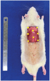
Figure 1.
Figure 1.
Surgical technique. A cutaneous lesion was induced in the dorsal region between the two scapulae with the largest axis in the longitudinal direction. Mononuclear stem cells were administered subcutaneously at eight points (yellow) along the margins and in the center of the wound.
Figure 2.
Figure 2.
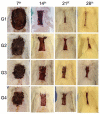
Macroscopic evaluation of wounds. Evaluation of wounds on the 7th, 14th, 21st, and 28th postoperative days for the groups G1 (control), G2 (SC), G3 (AM), and G4 (SC + AM). In the groups that received SC and/or AM, the wounds were significantly smaller after the initial 7 days of the repair process. After 28 days, the wounds in the control group were more contracted than those in the other groups.
Figure 3.
Figure 3.
Graph showing the contraction rate of the groups in the 7th, 14th, 21st, 28th days after surgery.
Figure 4.
Figure 4.
Photomicrography showing the area of collagen type I and III (400x). Picrossirius Red stains type I collagen in red and type III collagen in green, indicated by the solid yellow and dotted arrows, respectively. Analysis of captured images the polarized light microscope showed a predominance of collagen type I (mature) in all groups at the end of the experiments (28th day).
Figure 5.
Figure 5.
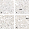
Photomicrography showing the area of elastic fiber (400x). Black arrows indicate elastic fibers stained black by the Verhoeff technique. After 28 days of the experiments, there was a predominance of elastic fibers in the AM group, followed by the SC + AM, SC and control groups.
Figure 6.
Figure 6.
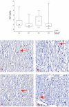
Comparison of groups in relation to the MMP-8. In the analysis of collagen remodeling on the 28th day, the results showed a lower expression of MMP-8 in the SC and SC + AM groups. Red arrows indicate MMP-8 expression stained brown (400X).
Figure 7.
Figure 7.
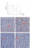
Comparison of groups in relation to the TGF-β. The expression of TGF-β showed the lowest percentages in the SC and SC + AM groups on the 28th day of the experiments. Red arrows indicate TGF-β expression stained brown (400X).
Figure 8.
Figure 8.
Comparison of groups in relation to the α-SMA. The expression of α-SMA verified the quantification of myofibroblasts and blood vessels, and showed the lowest percentages in the SC + AM group, followed by the AM, Control and SC group at the end of the experiments (28th day). Red arrows indicate expression of α-SMA stained brown (400X).

References

    1. Boyko TV, Longaker MT, Yang GP.. Laboratory models for the study of normal and pathologic wound healing. Plast Reconstr Surg. 2017;139:1–15. doi:10.1097/PRS.0000000000003077. - DOI - PubMed
    1. Brasil, Ministério da Saúde . Secretaria de Políticas de Saúde. Departamento de Atenção Básica. Manual de condutas para úlceras neurotróficas e traumáticas/Ministério da Saúde, Secretaria de Políticas de Saúde. Departamento de Atenção Básica. Brasília, Distrito Federal / Brazil: Ministério da Saúde, 2002. [accessed 2020 July]. http://bvsms.saude.gov.br/bvs/publicacoes/manual_feridas_final.pdf
    1. Broughton G, Janis JE, Attinger CE.. The basic science of wound healing. Plast Reconstr Surg. 2006;117:12S–34S. doi:10.1097/01.prs.0000225430.42531.c2. - DOI - PubMed
    1. Eming SA, Martin P, Tomic-Canic M. Wound repair and regeneration: mechanisms, signaling, and translation. Sci Transl Med. 2014;6:265sr6. doi:10.1126/scitranslmed.3009337. - DOI - PMC - PubMed
    1. Lombardi F, Palumbo P, Augello FR, Cifone MG, Cinque B, Giuliani M. Secretome of adipose tissue-derived stem cells (ASCs) as a novel trend in chronic non-healing wounds: an overview of experimental in vitro and in vivo studies and methodological variables. Int J Mol Sci. 2019;20:3721. doi:10.3390/ijms20153721. - DOI - PMC - PubMed

LinkOut - more resources