. 2021 Nov;20(11):e13494.
doi: 10.1111/acel.13494. Epub 2021 Oct 6.

Aging impairs human bone marrow function and cardiac repair following myocardial infarction in a humanized chimeric mouse

Affiliations

Aging impairs human bone marrow function and cardiac repair following myocardial infarction in a humanized chimeric mouse

Tina B Marvasti et al. Aging Cell. 2021 Nov.

Abstract

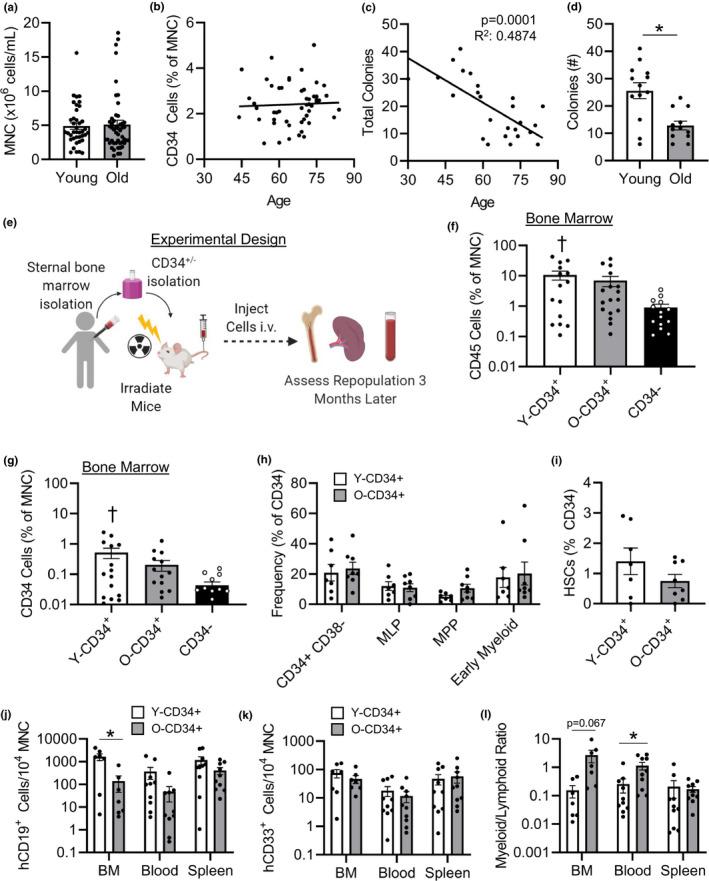
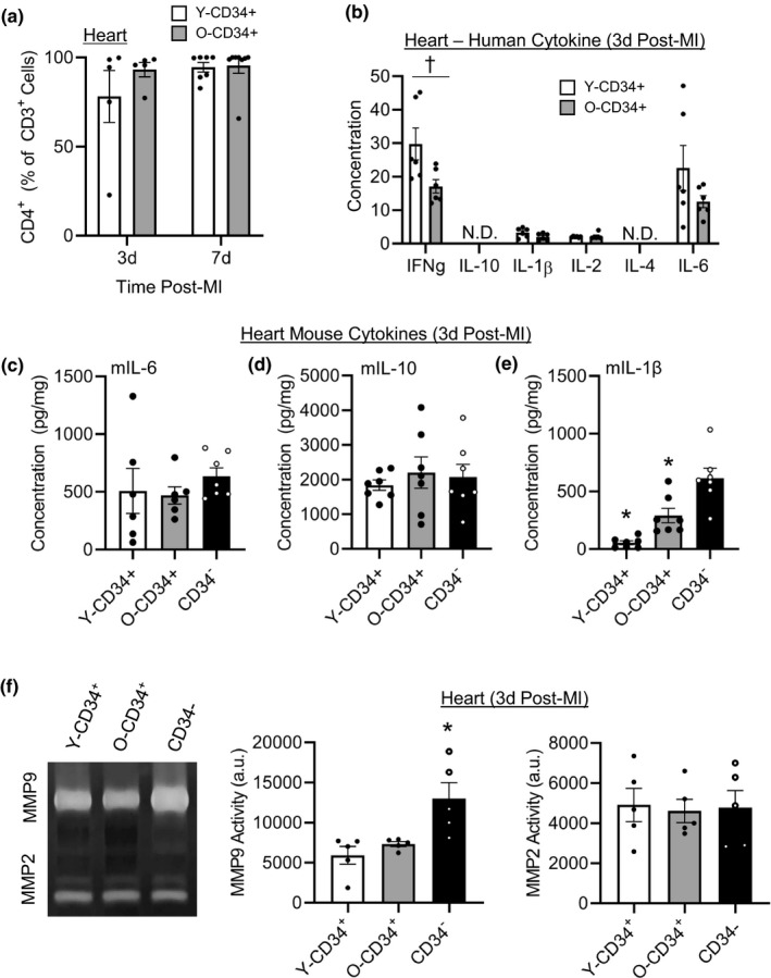
Ventricular remodeling following myocardial infarction (MI) is a major cause of heart failure, a condition prevalent in older individuals. Following MI, immune cells are mobilized to the myocardium from peripheral lymphoid organs and play an active role in orchestrating repair. While the effect of aging on mouse bone marrow (BM) has been studied, less is known about how aging affects human BM cells and their ability to regulate repair processes. In this study, we investigate the effect aging has on human BM cell responses post-MI using a humanized chimeric mouse model. BM samples were collected from middle aged (mean age 56.4 ± 0.97) and old (mean age 72.7 ± 0.59) patients undergoing cardiac surgery, CD34+/- cells were isolated, and NOD-scid-IL2rγnull (NSG) mice were reconstituted. Three months following reconstitution, the animals were examined at baseline or subjected to coronary artery ligation (MI). Younger patient cells exhibited greater repopulation capacity in the BM, blood, and spleen as well as greater lymphoid cell production. Following MI, CD34+ cell age impacted donor and host cellular responses. Mice reconstituted with younger CD34+ cells exhibited greater human CD45+ recruitment to the heart compared to mice reconstituted with old cells. Increased cellular responses were primarily driven by T-cell recruitment, and these changes corresponded with greater human IFNy levels and reduced mouse IL-1β in the heart. Age-dependent changes in BM function led to significantly lower survival, increased infarct expansion, impaired host cell responses, and reduced function by 4w post-MI. In contrast, younger CD34+ cells helped to limit remodeling and preserve function post-MI.

Keywords: aging; bone marrow transplant; humanized mice; myocardial infarction.


Conflict of interest statement

None.

Figures

FIGURE 1
FIGURE 1
Old CD34+ cells exhibit reduced colony formation and lower lymphoid cell repopulation following transplant. (a) Mononuclear cell concentration (n = 40, 56.4 ± 0.97 years old [Y‐CD34+], and n = 48, 72.7 ± 0.59 years old [O‐CD34+]) and (b) CD34+ cell frequency in younger and old patient BM (n = 51, 56.8 ± 1.2 years old [Y‐CD34+] vs. 73 ± 0.7 years old [O‐CD34+]). (c) Colony‐forming ability of CD34+ cells from younger and old patient BM (n = 25, 52.7 ± 2.5 years old [Y‐CD34+] vs. 75.1 ± 1.4 years old [O‐CD34+]). (d) And total number of colonies formed stratified based on age group (n = 13, 52.7 ± 2.5 years old [Y‐CD34+] and n = 12, 75.1 ± 1.4 years old [O‐CD34+]). (e) Schematic representation of experimental design for reconstitution model. (f) hCD45+, and (g) CD34+ frequency in the BM at 3 months postreconstitution (n = 15, 56.2 ± 1.6 years old [Y‐CD34+], n = 17, 71.8 ± 0.7 years old [O‐CD34+], n = 14, 68 ± 2.6 years old [CD34]). (h) Engrafted human hematopoietic progenitor cell frequencies in the BM of NSG mice transplanted with younger and old CD34+ bone marrow cells and (i) engrafted BM human HSCs 3 months after reconstitution (n = 7, 56.9 ± 1.4 years old [Y‐CD34+], n = 8, 73.1 ± 1.5 years old [O‐CD34+]). (j) Mice transplanted with younger CD34+ cells exhibited greater levels of B cells (CD19+) in the bone marrow 3 months post‐transplant; (k) myeloid cell frequency (CD33+) was similar between mice engrafted with younger and old cells and (l) myeloid (CD33+):lymphoid (CD19+) in the BM, blood, and spleen 3 months post‐reconstitution (n = 7–10/group, 57.6 ± 1.5 years old [Y‐CD34+] vs. 73.1 ± 1years old [O‐CD34+]).*p < 0.05 vs. indicated groups, p < 0.05 vs. CD34. Values are mean ± SEM
FIGURE 2
FIGURE 2
Age and cell type affect the long‐term functional outcome post‐MI. (a) Schematic representation of cardiac function analysis after reconstitution and coronary ligation. (b) Survival curve of Y/O‐CD34+ mice versus CD34 mice by 4 weeks post‐MI, *p < 0.05 Y‐CD34+ vs. all groups. (c) Functional analysis post‐MI measured by echocardiography demonstrating dimensions at (c) diastole and (d) systole and cardiac function demonstrated by (e) ejection fraction and (f) fractional shortening at 4 weeks post‐MI (55.3 ± 2.1 years old [Y‐CD34+] vs. 74.1 ± 1.7 years old [O‐CD34+], n = 7‐10/group). All timepoint values are provided in Table S1. (g) Gross tissue and (h) histological analyses with trichrome staining at 4 weeks post‐MI for analysis of scar size. Two different stained hearts are shown for each group. (i) Quantification of infarct expansion comparison (53.6 ± 2.5 years old [Y‐CD34+] vs.73 ± 0.6 years old [O‐CD34+], n = 4‐5/group). *p < 0.05 vs. Y‐CD34+ all groups at same time point, p < 0.05 O‐CD34+ vs. CD34 at same time point, and **p < 0.05 Y‐CD34+ vs. CD34 at same time point. Values are mean ± SEM
FIGURE 3
FIGURE 3
Younger CD34+ cells reduce remodeling and improve scar angiogenesis by 4 weeks post‐MI. (a) WGA staining of younger CD34+, old CD34+, and CD34 myocytes for cross‐sectional area analysis at 4w post‐MI, and quantification (52.2 ± 3.0 years old [YCD34+] vs. 71.2 ± 1.6 years old [O‐CD34+], vs. 59.2 ± 2.9 years old [CD34‐], n = 4/group). (b) Isolectin‐IB4 staining of the infarct region. (c) Mouse CD31+ cell staining in the infarct regions. (d) Quantification of isolectin staining for comparison between the three groups (50.6 ± 2.8 years old [Y‐CD34+] vs. 70.8 ± 1.3 years old [O‐CD34+], vs. 57.4 ± 2.1 years old [CD34‐], n = 5/group). (e) Quantification of mCD31 cell staining for angiogenesis comparison between the groups (50.6 ± 2.8 years old [Y‐CD34+] vs. 70.8 ± 1.3 years old [O‐CD34+], n = 5/group). (f) Human CD45+ staining demonstrating inflammatory cell infiltration into the myocardial infarct region at 3d post‐MI. (g) Quantification of the hCD45+ staining at 3d post‐MI among the younger CD34+ (50.6 ± 2.8 years old), old CD34+ (70.8 ± 1.3 years old), and CD34 hearts (n = 5/group). *p < 0.05 vs. all groups, p < 0.05 vs. CD34. Values are mean ± SEM
FIGURE 4
FIGURE 4
Aged CD34+ cells exhibit impaired human cell recruitment to the heart post‐MI. (a) Human CD45 cells (hCD45), (b) human T cells (hCD3), and (c) human myeloid (hCD33) cells in the mouse myocardium at baseline, 3, and 7 d post‐MI as measured by flow cytometry (56.5 ± 3.8 years old [Y‐CD34+] vs. 72.2 ± 0.8 years old [O‐CD34+] at baseline, 57 ± 1.2 years old [Y‐CD34+] vs. 71.9 ± 1.0 years old [O‐CD34+] at 3 d and 55.4 ± 1.7 years old [Y‐CD34+] vs. 72.7 ± 1.2 years old [O‐CD34+] at 7 d post‐MI, n = 4–10/group). (d) Representative flow cytometry plots showing differences in T‐cell cell number. (e) Circulatory hCD45+, (f) hCD3+, (g) CD33+ cell frequency in the blood of Y/O‐CD34+ reconstituted mice at baseline, 3 and 7 d post‐MI (56.5 ± 1.6 years old [Y‐CD34+] vs. 73.1 ± 1.0 years old [O‐CD34+] at baseline, 56.8 ± 0.7 years old [Y‐CD34+] vs. 72.2 ± 1.3 years old [O‐CD34+] at 3 d, and 58 ± 1.4 years old [Y‐CD34+] vs. 72.4 ± 1.1 years old [O‐CD34+] at 7 d post‐MI, n = 7–10/group). (h) Spleen hCD3+ cell frequency in chimeric mice at baseline, 3 and 7 d post‐MI (56.5 ± 1.6 years old [Y‐CD34+] vs. 73.1 ± 1.0 years old [OCD34+] at baseline, 56.5 ± 0.7 years old [Y‐CD34+] vs. 72.2 ± 1.3 years old [O‐CD34+] at 3 d, and 58.4 ± 1.4 years old [Y‐CD34+] vs. 72 ± 1.1 years old [O‐CD34+] at 7 d post‐MI, n = 7–10/group). *p < 0.05 between groups at indicated time point. Values are mean ± SEM
FIGURE 5
FIGURE 5
CD34+ cell age affects early mediators of cardiac repair. (a) T‐cell subtype analysis of CD4+ frequency 3 and 7 d post‐MI (58.2 ± 1.9 years old [Y‐CD34+] vs. 71.9 ± 1.0 years old [O‐CD34+] at 3 d and 57 ± 1.6 years old [Y‐CD34+] vs. 73 ± 0.9 years old [OCD34+] at 7 d post‐MI, n = 5–8/group). (b) Human cytokine analysis of pro‐ and anti‐inflammatory cytokines in younger and older CD34+ hearts at 3 d post‐MI (56 ± 2.1 years old [Y‐CD34+] vs. 69.5 ± 1.7 years old [O‐CD34+], n = 6/group). Myocardial mouse cytokines (c) IL‐6, (d) IL‐10, and (e) IL‐1β in Y/O CD34+ and CD34 myocardium (56 ± 2.1 years old [Y‐CD34+] vs. 69.5 ± 1.7 years old [O‐CD34+], n = 7/group). (f) MMP2 and MMP9 activity at 3 d post‐MI in in Y/O CD34+ and CD34 infarcted myocardium measured using zymography (55.6 ± 2.2 years old [Y‐CD34+] vs. 69.8 ± 1.9 years old [O‐CD34+], n = 5/group). *p < 0.05 vs all other groups, p < 0.05 groups as indicated. Values are mean ± SEM
FIGURE 6
FIGURE 6
CD34+ cell age alters the infiltration of mouse (host) myeloid cells post‐MI. (a) Quantification of mouse CD45+ (mCD45) cells in the heart 3‐ and 7 d post‐MI (56.3 ± 1.1 years old [Y‐CD34+] vs. 70.1 ± 1.6 years old [O‐CD34+], vs. 68.5 ± 3.5 years old [CD34‐] at 3 d and 61.5 ± 1.4 years old [Y‐CD34+] vs. 70.3 ± 1.4 years old [O‐CD34+], vs. 65.5 ± 3.1 years old [CD34‐] at 7 d, n = 3–7/group). Quantification of mouse (b) neutrophil, (c) Ly6Chi monocytes, and (d) Ly6Cneg monocytes 3 and 7 d post‐MI in Y‐CD34+, O‐CD34+, CD34, and WT NSG mice (n = 3–7/group). (e) Quantification of total macrophages in heart 3‐ and 7‐d post‐MI and (f) frequency of CD206+ macrophages in the heart at 3 d post‐MI. p < 0.05 vs. CD34 at the same timepoint, †† p < 0.05 vs. WT NSG at the same timepoint, *p < 0.05 vs indicated groups at same timepoint, €p < 0.05 vs. O‐CD34+ at the same timepoint. Values are mean ± SEM

References

    1. Alibhai, F. J. , & Li, R. K. (2020). Targeting aged bone marrow for systemic rejuvenation. Aging, 12(3), 2024–2025. 10.18632/aging.102838 - DOI - PMC - PubMed
    1. Alibhai, F. J. , Lim, F. , Yeganeh, A. , DiStefano, P. V. , Binesh‐Marvasti, T. , Belfiore, A. , Wlodarek, L. , Gustafson, D. , Millar, S. , Li, S.‐H. , Weisel, R. D. , Fish, J. E. , & Li, R.‐K. (2020). Cellular senescence contributes to age‐dependent changes in circulating extracellular vesicle cargo and function. Aging Cell, 19(3), e13103. 10.1111/acel.13103 - DOI - PMC - PubMed
    1. Broxmeyer, H. E. , Liu, Y. , Kapur, R. , Orschell, C. M. , Aljoufi, A. , Ropa, J. P. , Trinh, T. , Burns, S. , & Capitano, M. L. (2020). Fate of hematopoiesis during aging. What do we really know, and what are its implications? Stem Cell Reviews and Reports, 16(6), 1020–1048. 10.1007/s12015-020-10065-y - DOI - PMC - PubMed
    1. Cheng, X. , Liao, Y. H. , Ge, H. , Li, B. , Zhang, J. , Yuan, J. , & Zhang, L. (2005). TH1/TH2 functional imbalance after acute myocardial infarction: coronary arterial inflammation or myocardial inflammation. Journal of Clinical Immunology, 25(3), 246–253. 10.1007/s10875-005-40880 - DOI - PubMed
    1. Cooper, L. B. , DeVore, A. D. , & Michael Felker, G. (2015). The impact of worsening heart failure in the United States. Heart Failure Clinics, 11(4), 603–614. 10.1016/j.hfc.2015.07.004 - DOI - PMC - PubMed

Publication types

Grants and funding