. 2021 Nov;25(21):10028-10038.
doi: 10.1111/jcmm.16913. Epub 2021 Oct 6.

A comparative analysis of deferoxamine treatment modalities for dermal radiation-induced fibrosis

Affiliations

A comparative analysis of deferoxamine treatment modalities for dermal radiation-induced fibrosis

Christopher V Lavin et al. J Cell Mol Med. 2021 Nov.

Abstract

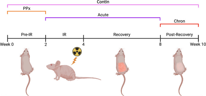
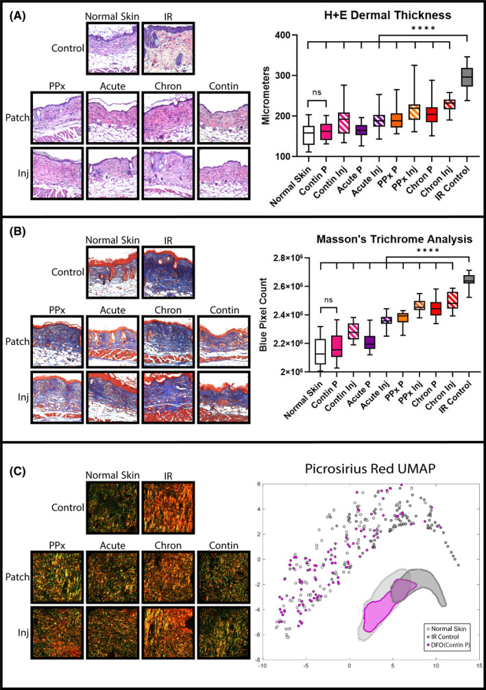
The iron chelator, deferoxamine (DFO), has been shown to potentially improve dermal radiation-induced fibrosis (RIF) in mice through increased angiogenesis and reduced oxidative damage. This preclinical study evaluated the efficacy of two DFO administration modalities, transdermal delivery and direct injection, as well as temporal treatment strategies in relation to radiation therapy to address collateral soft tissue fibrosis. The dorsum of CD-1 nude mice received 30 Gy radiation, and DFO (3 mg) was administered daily via patch or injection. Treatment regimens were prophylactic, during acute recovery, post-recovery, or continuously throughout the experiment (n = 5 per condition). Measures included ROS-detection, histology, biomechanics and vascularity changes. Compared with irradiated control skin, DFO treatment decreased oxidative damage, dermal thickness and collagen content, and increased skin elasticity and vascularity. Metrics of improvement in irradiated skin were most pronounced with continuous transdermal delivery of DFO. In summary, DFO administration reduces dermal fibrosis induced by radiation. Although both treatment modalities were efficacious, the transdermal delivery showed greater effect than injection for each temporal treatment strategy. Interestingly, the continuous patch group was more similar to normal skin than to irradiated control skin by most measures, highlighting a promising approach to address detrimental collateral soft tissue injury following radiation therapy.

Keywords: dermal fibrosis; fibrosis treatment; radiation therapy; translational science.


Conflict of interest statement

Dr. Michael Longaker and Dr. Geoffrey Gurtner have equity stakes in TauTona Group, the DFO patch supplier. Dr. Geoffrey Gurtner holds a patent for topical and transdermal HIF‐1 modulators for chronic wound treatment. Dr. Derrick Wan, Dr. Michael Longaker and Dr. Geoffrey Gurtner hold a patent for the use of DFO in conditioning irradiated tissue.

Figures

FIGURE 1
FIGURE 1
Treatment regimen schematic. Treated mice received DFO via patch or injection; both administration routes were tested within each treatment period (PPx, Acute, Chron, Contin). Abbreviations: Chron, chronic; Contin, continuous; DFO, deferoxamine; IR, irradiation; PPx, prophylactic
FIGURE 2
FIGURE 2
ROS and oxidative stress markers. (A) Perls Prussian Blue staining for ferric iron: representative 40× images (left) and quantification (right chart). DFO treatment decreased mean free iron in the dermis compared with IR Control and Normal Skin (*p < 0.05, **p < 0.01, ****p < 0.0001). (B) Representative 20× 8‐Iso images (left) and quantification (right chart). DFO treatments decreased ROS‐mediated lipid peroxidation compared with IR Control skin (****p < 0.0001). (C) ELISA of glutathione oxidation state (left chart) and BAX protein levels (right chart). DFO treatment decreased ratio of GSSG:GSH compared with IR Control skin (****p < 0.0001). DFO treatment also decreased BAX apoptotic protein level compared with IR Control skin (****p < 0.0001). Greater reduction in GSSG:GSH and BAX protein for DFO Patch relative to Injection were noted. Abbreviations: 8‐Iso, 8‐Isoprostane; BAX, Bcl‐2‐associated protein X; DFO, deferoxamine; ELISA, enzyme‐linked immunosorbent assay; GSSG:GSH, ratio of oxidized to reduced glutathione; Inj, injection; ns, not significant; ROS, reactive oxygen species
FIGURE 3
FIGURE 3
Histological RIF quantification. (A) Representative 20× H + E specimens (left) and quantification of dermal thickness (right chart). Mean dermal thickness was reduced with DFO treatment compared with IR control skin (****p < 0.0001). Greatest reduction in dermal thickness with continuous patch (Contin P) was noted, which was similar to normal skin. (B) Representative 20× TC samples (left) and quantification of collagen content (right chart). Mean collagen content was reduced with DFO treatment compared with IR Control skin (****p < 0.0001). Greatest reduction in collagen content with continuous patch (Contin P) was noted, which was similar to normal skin. (C) Representative 40× Picro images (left) and machine‐learning algorithm‐derived collagen ultrastructure UMAP representation of extracellular matrix characteristics for normal skin (light grey), IR Control (dark grey), and continuous DFO patch (pink). Normal Skin and Contin P DFO treatment overlapped more with each other than with IR Control skin, represented by cluster shape approximation underneath plot points. Abbreviations: Chron, chronic; Contin, continuous; DFO, deferoxamine; H+E, haematoxylin and eosin; Inj, injection; IR, irradiation; P, patch; Picro, Picrosirius Red; PPx, prophylactic; RIF, radiation‐induced fibrosis; TC, Masson's Trichrome; UMAP, uniform manifold approximation and projection
FIGURE 4
FIGURE 4
Biomechanical properties. (A) Representative suction Cutometer curves at week 10. One curve displayed for each group. (B) Elasticity analysis at week 10. Mean maximum suction amplitude was significantly higher for DFO treated groups compared with IR Control skin (****p < 0.0001), with greatest amplitude appreciated for continuous DFO patch treatment (Contin P). (C) Post‐harvest tensile testing revealed most DFO treatment groups were significantly less stiff than IR Control skin. Patch or injection treatment during post‐recovery chronic phase (Chron P and Chron Inj) and DFO injection during acute recovery (Acute Inj) groups had lower mean Young's Moduli vs. IR Control, however, none of these three groups achieved statistical significance like the other groups (**p < 0.01, ****p < 0.0001). Abbreviations: Chron, chronic; Contin, continuous; DFO, deferoxamine; Inj, injection; IR, irradiation; ns, not significant; P, patch; PPx, prophylactic
FIGURE 5
FIGURE 5
Dermal microvasculature analysis. (A) Laser Doppler perfusion imaging representative heat maps of dorsal skin at week 10 (left) and longitudinal perfusion tracking measured biweekly throughout experiment (right chart). (B) Mean perfusion analysis at week 4 (left chart) and week 10 (right chart). At week 4, immediate radiation injury elevated mean perfusion for all groups compared with Normal Skin (****p < 0.0001). By week 10, DFO treatment groups' mean perfusion levels decreased less than IR Control skin (***p < 0.001, ****p < 0.0001). The highest perfusion measured with continuous DFO patch treatment (Contin P) was noted, which was similar to normal skin. (C) Representative 20× CD31 immunofluorescent images (left) and quantification (right). Most DFO treatments increased immunofluorescent staining for CD31 compared with IR Control skin (**p < 0.01, ***p < 0.001, ****p < 0.0001). Note that, prophylactic DFO patch (PPx P) and prophylactic DFO injection (PPx Inj) groups' mean red pixel area was greater than IR Control; however, neither achieved significance. Abbreviations: Chron, chronic; Contin, continuous; DFO, deferoxamine; Inj, injection; IR, irradiation; ns, not significant; P, patch; PPx, prophylactic

References

    1. Bentzen SM. Preventing or reducing late side effects of radiation therapy: radiobiology meets molecular pathology. Nat Rev Cancer. 2006;6(9):702‐713. doi: 10.1038/nrc1950 - DOI - PubMed
    1. Bray FN, Simmons BJ, Wolfson AH, Nouri K. Acute and chronic cutaneous reactions to ionizing radiation therapy. Dermatol Ther. 2016;6(2):185‐206. doi: 10.1007/s13555-016-0120-y - DOI - PMC - PubMed
    1. Salvo N, Barnes E, van Draanen J, et al. Prophylaxis and management of acute radiation‐induced skin reactions: a systematic review of the literature. Curr Oncol. 2010;17(4):94‐112. doi: 10.3747/co.v17i4.493 - DOI - PMC - PubMed
    1. Baskar R, Lee KA, Yeo R, Yeoh K‐W. Cancer and radiation therapy: current advances and future directions. Int J Med Sci. 2012;9(3):193‐199. doi: 10.7150/ijms.3635 - DOI - PMC - PubMed
    1. Delaney G, Jacob S, Featherstone C, Barton M. The role of radiotherapy in cancer treatment: estimating optimal utilization from a review of evidence‐based clinical guidelines. Cancer. 2005;104(6):1129‐1137. doi: 10.1002/cncr.21324 - DOI - PubMed

Publication types