. 2021 Oct 27;6(5):e0067621.
doi: 10.1128/mSphere.00676-21. Epub 2021 Oct 6.

Temperature Influences the Composition and Cytotoxicity of Extracellular Vesicles in Staphylococcus aureus

Affiliations

Temperature Influences the Composition and Cytotoxicity of Extracellular Vesicles in Staphylococcus aureus

Paul Briaud et al. mSphere. .

Abstract

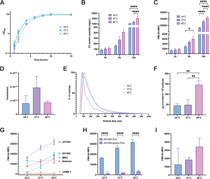
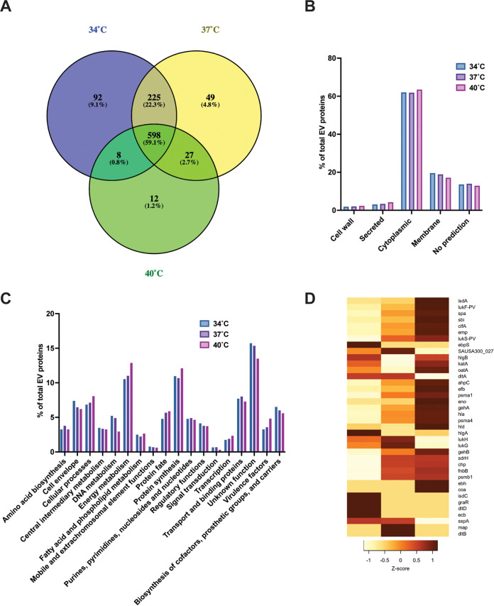
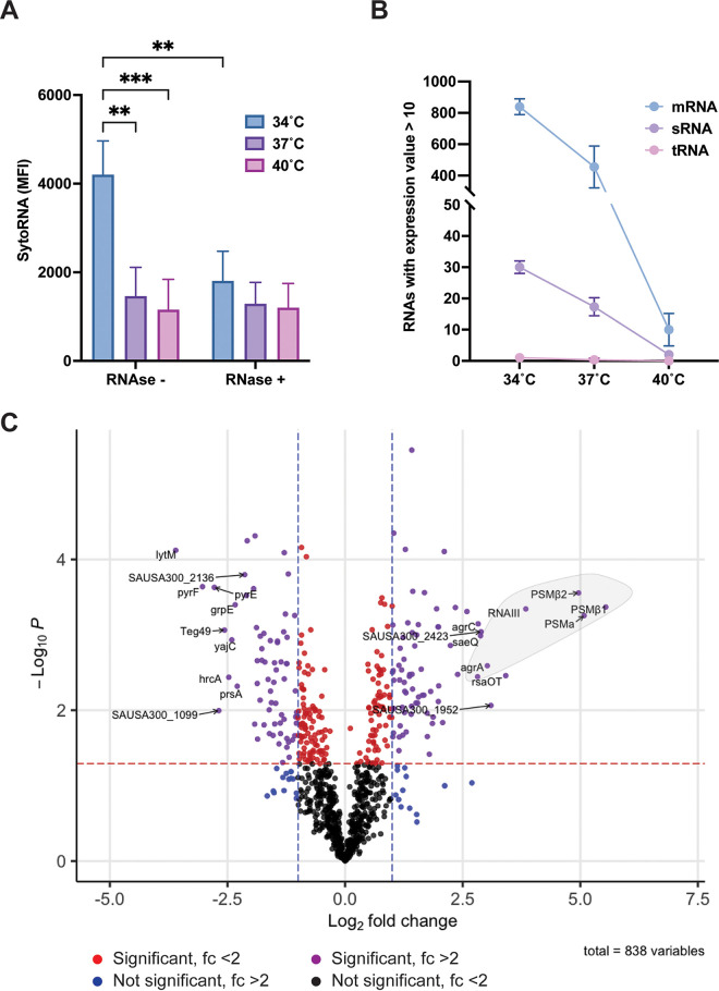
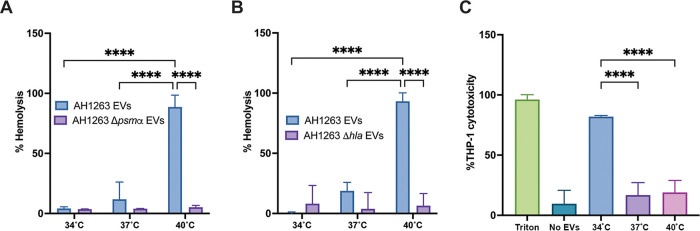
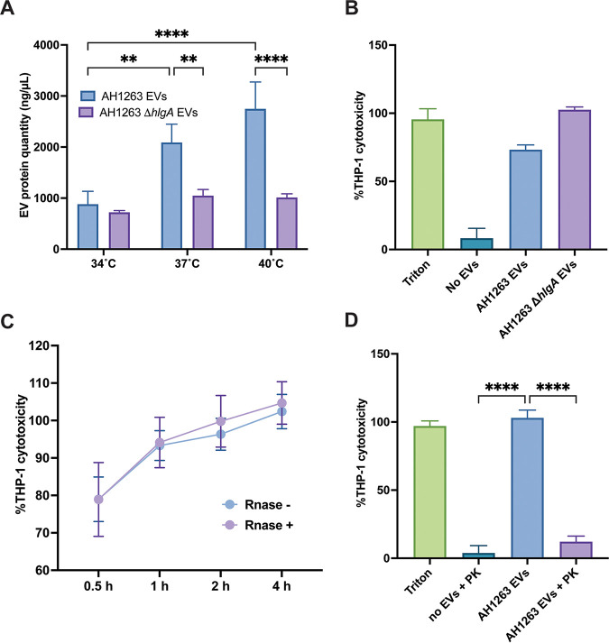
Staphylococcus aureus is a pathogenic bacterium but also a commensal of skin and anterior nares in humans. As S. aureus transits from skins/nares to inside the human body, it experiences changes in temperature. The production and content of S. aureus extracellular vesicles (EVs) have been increasingly studied over the past few years, and EVs are increasingly being recognized as important to the infectious process. Nonetheless, the impact of temperature variation on S. aureus EVs has not been studied in detail, as most reports that investigate EV cargoes and host cell interactions are performed using vesicles produced at 37°C. Here, we report that EVs in S. aureus differ in size and protein/RNA cargo depending on the growth temperature used. We demonstrate that the temperature-dependent regulation of vesicle production in S. aureus is mediated by the alpha phenol-soluble modulin peptides (αPSMs). Through proteomic analysis, we observed increased packaging of virulence factors at 40°C, whereas the EV proteome has greater diversity at 34°C. Similar to the protein content, we perform transcriptomic analysis and demonstrate that the RNA cargo also is impacted by temperature. Finally, we demonstrate greater αPSM- and alpha-toxin-mediated erythrocyte lysis with 40°C EVs, but 34°C EVs are more cytotoxic toward THP-1 cells. Together, our study demonstrates that small temperature variations have great impact on EV biogenesis and shape the interaction with host cells. IMPORTANCE Extracellular vesicles (EVs) are lipid bilayer spheres that contain proteins, nucleic acids, and lipids secreted by bacteria. They are involved in Staphylococcus aureus infections, as they package virulence factors and deliver their contents inside host cells. The impact of temperature variations experienced by S. aureus during the infectious process on EVs is unknown. Here, we demonstrate the importance of temperature in vesicle production and packaging. High temperatures promote packaging of virulence factors and increase the protein and lipid concentration but reduce the overall RNA abundance and protein diversity in EVs. The importance of temperature changes is highlighted by the fact that EVs produced at low temperature are more toxic toward macrophages, whereas EVs produced at high temperature display more hemolysis toward erythrocytes. Our research brings new insights into temperature-dependent vesiculation and interaction with the host during S. aureus transition from colonization to virulence.

Keywords: Staphylococcus aureus; extracellular vesicle; membrane vesicle; proteomics; temperature; transcriptomics.


Figures

FIG 1
FIG 1
Temperature-dependent EV production in S. aureus. (A) Growth curves of wild-type AH1263 over 15 h in TSB at 34°C, 37°C, or 40°C. (B and C) Vesicles produced from AH1263 at 3 h, 6 h, and 15 h were analyzed for both protein (B) and lipid concentration (C). (D to F) Particle concentration (D) and size distribution (E) of S. aureus EV preparations were analyzed by NTA, and the relative concentration of proteins were estimated (F). (G) Vesicle production at 34°C, 37°C, and 40°C in different wild-type strains of S. aureus was analyzed by FM4-64. (H) EVs from the Δpsmα mutant were analyzed and compared to AH1263 EVs. The level of EV lipid from the Δpsmα mutant is indicated in more detail in panel I. Data represent means ± standard deviations (SD) from 3 independent experiments. *, P < 0.05; **, P < 0.01; ***, P < 0.001; ****, P < 0.0001, as calculated by analysis of variance (ANOVA) according to Materials and Methods.
FIG 2
FIG 2
Temperature impacts S. aureus EV protein content. A mass spectrometry analysis was conducted on AH1263 EVs produced at 34°C, 37°C, and 40°C at 15 h. (A) Venn-diagram of EV proteins at each temperature. (B) EV protein distribution sorted by their predicted localization by PsortB. (C) Analysis and distribution of EV proteins by functional categories. (D) Heatmap depicting the Z-score of select virulence factors detected in EVs. A dark red color indicates a high abundance, whereas a light color depicts low abundance in EVs.
FIG 3
FIG 3
Temperature influences EV RNA composition. (A) The Syto RNAselect dye was used to quantify the amount of total RNA (surface and lumen associated; RNase) or exclusively lumen-associated RNA (RNase+) in AH1263 EVs produced at 15 h at 34, 37, or 40°C. Data represent means ± SD from 3 independent experiments. **, P < 0.01; ***, P < 0.001, as calculated by ANOVA according to Materials and Methods. MFI, mean fluorescence intensity. (B) RNAs were sorted according to their classes in the cell (mRNA, sRNA, and tRNA). Only transcripts with mean expression values of >10 and >80% of unique mapped reads were used. (C) Volcano plot of differential expression analysis between 34°C and 37°C EV RNAs. Purple dots depict significant changes with log2 fold change of >1 and –log10P value of >1.3 (P < 0.05) cutoffs. Red and blue dots show transcripts reaching either significant P value (red dotted line) or log2 fold change (blue dotted lines) thresholds. Black dots indicate transcripts not differentially present. The first 10 differentially present transcripts under each condition are indicated. The bright gray circle points out the transcripts regulated by the Agr regulon.
FIG 4
FIG 4
EV cytotoxicity is temperature dependent. Human (A) or rabbit (B) erythrocyte hemolysis was assessed by incubating 20 μg of EVs produced at each temperature for either 15 min (for human blood) or 2.5 min (for rabbit blood) at 37°C. The hemolytic activity was determined by reading the absorbance of the samples at OD543. (C) THP-1 cells were incubated with 5 μg of EVs for 4 h prior to cell viability assessment. Data represent means ± SD from 3 independent experiments. ****, P < 0.0001 as calculated by analysis of variance (ANOVA) according to Materials and Methods.
FIG 5
FIG 5
Surface-associated proteins are responsible for cytotoxicity of 34°C EVs. (A) Vesicles produced at 15 h from AH1263 WT and AH1263 carrying a transposon inserted into the hlgA gene (hlgA) were analyzed for protein content. (B to D) THP-1 cells were exposed to 5 μg of either hlgA mutant EVs (B) for 4 h, AH1263 EVs (with or without RNase) for 0.5 h, 1 h, 2 h, or 4 h (C), or proteinase K-treated EVs for 4 h (D) prior to cell viability assessment. Data represent means ± SD from 3 independent experiments. ****, P < 0.0001 as calculated by analysis of variance (ANOVA) according to Materials and Methods.

References

    1. Briaud P, Carroll RK. 2020. Extracellular-vesicle (EV) biogenesis and functions in Gram-positive bacteria. Infect Immun 88:e00433-20. doi: 10.1128/IAI.00433-20. - DOI - PMC - PubMed
    1. Jan AT. 2017. Outer membrane vesicles (OMVs) of Gram-negative bacteria: a perspective update. Front Microbiol 8:1053. doi: 10.3389/fmicb.2017.01053. - DOI - PMC - PubMed
    1. Schwechheimer C, Kuehn MJ. 2015. Outer-membrane vesicles from Gram-negative bacteria: biogenesis and functions. Nat Rev Microbiol 13:605–619. doi: 10.1038/nrmicro3525. - DOI - PMC - PubMed
    1. Brown L, Wolf JM, Prados-Rosales R, Casadevall A. 2015. Through the wall: extracellular vesicles in Gram-positive bacteria, mycobacteria and fungi. Nat Rev Microbiol 13:620–630. doi: 10.1038/nrmicro3480. - DOI - PMC - PubMed
    1. Lee J, Lee E-Y, Kim S-H, Kim D-K, Park K-S, Kim KP, Kim Y-K, Roh T-Y, Gho YS. 2013. Staphylococcus aureus extracellular vesicles carry biologically active β-lactamase. Antimicrob Agents Chemother 57:2589–2595. doi: 10.1128/AAC.00522-12. - DOI - PMC - PubMed

Publication types