Clinical Trial
. 2021 Aug 12;10(10):13.
doi: 10.1167/tvst.10.10.13.

One-Year Follow-Up in a Phase 1/2a Clinical Trial of an Allogeneic RPE Cell Bioengineered Implant for Advanced Dry Age-Related Macular Degeneration

Affiliations
Clinical Trial

One-Year Follow-Up in a Phase 1/2a Clinical Trial of an Allogeneic RPE Cell Bioengineered Implant for Advanced Dry Age-Related Macular Degeneration

Amir H Kashani et al. Transl Vis Sci Technol. .

Abstract

Purpose: To report 1-year follow-up of a phase 1/2a clinical trial testing a composite subretinal implant having polarized human embryonic stem cell (hESC)-derived retinal pigment epithelium (RPE) cells on an ultrathin parylene substrate in subjects with advanced non-neovascular age-related macular degeneration (NNAMD).

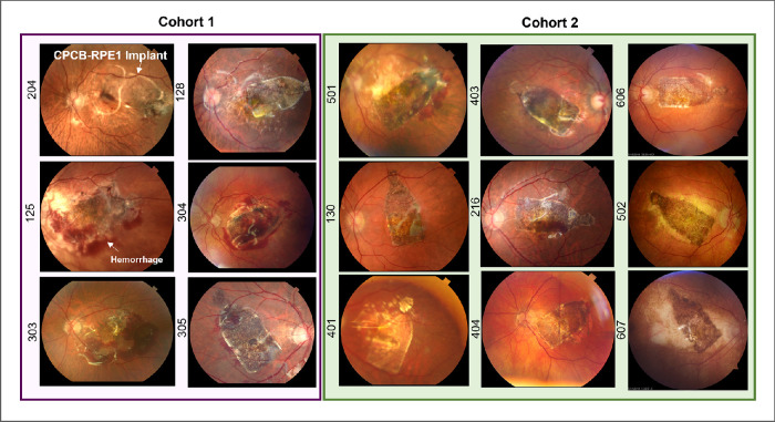
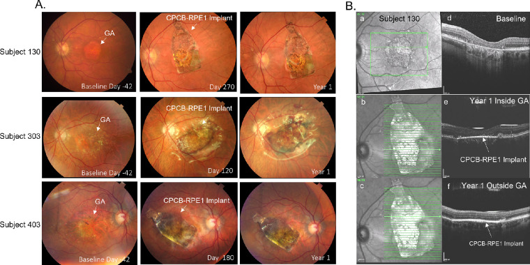
Methods: The phase 1/2a clinical trial included 16 subjects in two cohorts. The main endpoint was safety assessed at 365 days using ophthalmic and systemic exams. Pseudophakic subjects with geographic atrophy (GA) and severe vision loss were eligible. Low-dose tacrolimus immunosuppression was utilized for 68 days in the peri-implantation period. The implant was delivered to the worst seeing eye with a custom subretinal insertion device in an outpatient setting. A data safety monitoring committee reviewed all results.

Results: The treated eyes of all subjects were legally blind with a baseline best-corrected visual acuity (BCVA) of ≤ 20/200. There were no unexpected serious adverse events. Four subjects in cohort 1 had serious ocular adverse events, including retinal hemorrhage, edema, focal retinal detachment, or RPE detachment, which was mitigated in cohort 2 using improved hemostasis during surgery. Although this study was not powered to assess efficacy, treated eyes from four subjects showed an increased BCVA of >5 letters (6-13 letters). A larger proportion of treated eyes experienced a >5-letter gain when compared with the untreated eye (27% vs. 7%; P = not significant) and a larger proportion of nonimplanted eyes demonstrated a >5-letter loss (47% vs. 33%; P = not significant).

Conclusions: Outpatient delivery of the implant can be performed routinely. At 1 year, the implant is safe and well tolerated in subjects with advanced dry AMD.

Translational relevance: This work describes the first clinical trial, to our knowledge, of a novel implant for advanced dry AMD.


Conflict of interest statement

Disclosure: A.H. Kashani, Carl Zeiss Meditec (F); J.S. Lebkowski, Regenerative Patch Technologies (E, I, P); F.M. Rahhal, None; R.L. Avery, None; H. Salehi-Had, None; S. Chen, None; C. Chan, None; N. Palejwala, None; A. Ingram, Regenerative Patch Technologies (C); W. Dang, None; C.-M. Lin, None; D. Mitra, None; B.O. Pennington, Regenerative Patch Technologies (E, P); C. Hinman, Regenerative Patch Technologies (E); M.A. Faynus, Regenerative Patch Technologies (E); J.K. Bailey, Regenerative Patch Technologies (E); S. Mohan, None; N. Rao, None; L.V. Johnson, Regenerative Patch Technologies (E, I, P); D.O. Clegg, Regenerative Patch Technologies (I, C, P); D.R. Hinton, Regenerative Patch Technologies (I, C, P); M.S. Humayun, Regenerative Patch Technologies (I, C, P)

Figures

Figure 1.
Figure 1.
The CPCB-RPE1 implant. (A) The hESC-derived RPE cells seeded and cultured on the synthetic parylene membrane grow as a uniform monolayer of hexagonal, pigmented cells with significant similarity to RPE cells in vivo. (B) Scanning electron micrograph of the underside of the parylene membrane showing submicron-thick, circular regions (40-µm diameter) that facilitate diffusion of macromolecules and the supporting matrix. (C) Bright-field image of a single CPCB-RPE1 implant with pigmented, mature, and confluent RPE. The implant is 3.5 × 6.25 × 0.006 mm in size.
Figure 2.
Figure 2.
Color fundus photographs of all implanted subjects 28 days post-implantation. The left panel (purple box) illustrates all subjects enrolled in cohort 1. The right panel (green box) illustrates all subjects enrolled in cohort 2. Numbers on the left of each image denote the subject identifier. In all cases, the CPCB-RPE1 implant was successfully targeted to the subretinal space including the area of geographic atrophy and covering the majority of the lesion. Hemorrhage at day 28 was noted in four (subjects 125, 303, 304, and 305) of the six implanted subjects in cohort 1 and was reduced substantially at the same post-implantation time point in cohort 2.
Figure 3.
Figure 3.
Color fundus photographs and OCT images before and after implantation of CPCB-RPE1. (A) Color fundus photographs from subjects 130 (top row), 303 (middle row), and 403 (bottom row) at baseline, at an intermediate time point, and at 1 year post-implantation. (B) En face OCT images of subject 130 before implantation (a) and at 1 year post-implantation inside (b) and outside (c) the GA lesion. Corresponding B-scans through the area of geographic atrophy prior to implantation (d) and at 1 year post-implantation (e, f) show loss of RPE and photoreceptors prior to treatment. A bright hyperreflective line in the subretinal space indicates the location of the CPCB-RPE1 implant (e, f). The absence of hypertransmission defect in the post-implant images supports survival of transplanted RPE within the region of geographic atrophy.

References

    1. Kashani AH. Stem cell therapy in nonneovascular age-related macular degeneration. Invest Ophthalmol Vis Sci . 2016; 57(5): ORSFm1–ORSFm9. - PubMed
    1. Nazari H, Zhang L, Zhu D, et al. .. Stem cell based therapies for age-related macular degeneration: the promises and the challenges. Prog Retin Eye Res . 2015; 48: 1–39. - PubMed
    1. Miller JW. Age-related macular degeneration revisited–piecing the puzzle: the LXIX Edward Jackson memorial lecture. Am J Ophthalmol . 2013; 155(1): 1–35.e13. - PubMed
    1. Age-Related Eye Disease Study Research Group. A randomized, placebo-controlled, clinical trial of high-dose supplementation with vitamins C and E, beta carotene, and zinc for age-related macular degeneration and vision loss: AREDS report no. 8. Arch Ophthalmol . 2001; 119(10): 1417–1436. - PMC - PubMed
    1. Agrón E, Mares J, Clemons TE, et al. .. Dietary nutrient intake and progression to late age-related macular degeneration in the Age-Related Eye Disease studies 1 and 2. Ophthalmology. 2021; 128(3): 425–442. - PMC - PubMed

Publication types