Review
. 2022 Jan 20;34(1):103-128.
doi: 10.1093/plcell/koab249.

Cell biology of primary cell wall synthesis in plants

Affiliations
Review

Cell biology of primary cell wall synthesis in plants

Ying Gu et al. Plant Cell. .

Abstract

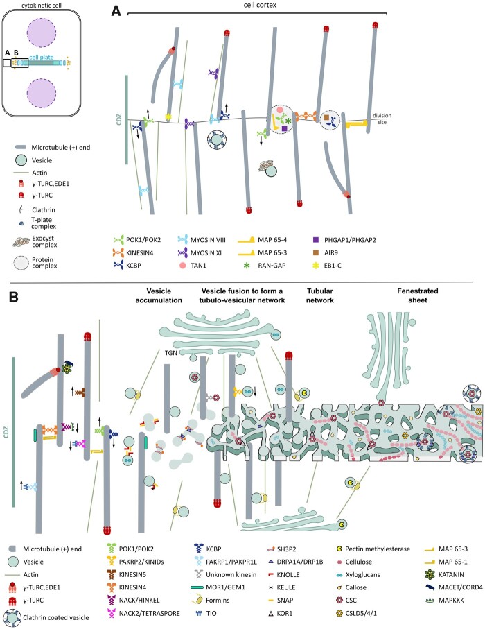
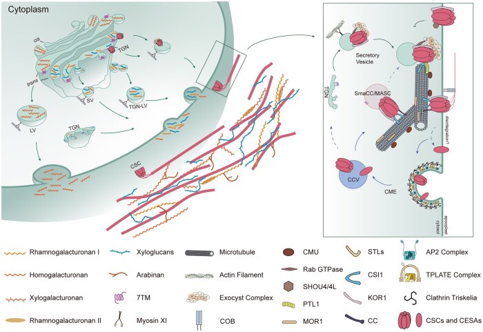
Building a complex structure such as the cell wall, with many individual parts that need to be assembled correctly from distinct sources within the cell, is a well-orchestrated process. Additional complexity is required to mediate dynamic responses to environmental and developmental cues. Enzymes, sugars, and other cell wall components are constantly and actively transported to and from the plasma membrane during diffuse growth. Cell wall components are transported in vesicles on cytoskeletal tracks composed of microtubules and actin filaments. Many of these components, and additional proteins, vesicles, and lipids are trafficked to and from the cell plate during cytokinesis. In this review, we first discuss how the cytoskeleton is initially organized to add new cell wall material or to build a new cell wall, focusing on similarities during these processes. Next, we discuss how polysaccharides and enzymes that build the cell wall are trafficked to the correct location by motor proteins and through other interactions with the cytoskeleton. Finally, we discuss some of the special features of newly formed cell walls generated during cytokinesis.


Figures

Figure 1
Figure 1
Model of land-plant cytokinesis. A, The cell cortex underlying the plasma membrane before it comes into contact with the phragmoplast. B, Phragmoplast, membrane, and cell wall structures that form the cell plate from left to right: vesicle accumulation, vesicle fusion to form a tubulo-vesicular network, a tubular network, and fenestrated sheet.
Figure 2
Figure 2
Model of trafficking of non-cellulosic polysaccharides and cellulose in the primary cell wall. The synthesis of non-cellulosic polysaccharides occurs in the Golgi. Non-cellulosic polysaccharides include xyloglucan and pectins. They are exported along actin filaments or microtubules in vesicles from the Golgi to the plasma membrane and incorporated into the apoplast. Cellulose is synthesized exclusively at the plasma membrane. The components involved in cellulose synthesis and its regulation are highlighted in the box on the right. Proteins associated with CSCs include Cellulose Synthase Interacting Protein 1 (CSI1), Companions of Cellulose synthase (CC), KORRIGAN1 (KOR1), and PATROL1 (PTL1). The accumulation of CSCs at the plasma membrane is influenced by exocytosis and endocytosis. Microtubules and actin filaments play important roles in the trafficking of CSCs to and from the membrane.

Comment in

  • Back to the roots: A focus on plant cell biology.
    Weijers D, Bezanilla M, Jiang L, Roeder AHK, Williams M. Weijers D, et al. Plant Cell. 2022 Jan 20;34(1):1-3. doi: 10.1093/plcell/koab278. Plant Cell. 2022. PMID: 34755878 Free PMC article. No abstract available.

References

    1. Abou-Saleh RH, Hernandez-Gomez MC, Amsbury S, Paniagua C, Bourdon M, Miyashima S, Helariutta Y, Fuller M, Budtova T, Connell SD, et al. (2018) Interactions between callose and cellulose revealed through the analysis of biopolymer mixtures. Nat Commun 9: 1–13 - PMC - PubMed
    1. Abu-Abied M, Belausov E, Hagay S, Peremyslov V, Dolja V, Sadot E (2018) Myosin XI-K is involved in root organogenesis, polar auxin transport, and cell division. J Exp Bot 69: 2869–2881 - PMC - PubMed
    1. Ahn G, Kim H, Kim DH, Hanh H, Yoon Y, Singaram I, Wijesinghe KJ, Johnson KA, Zhuang X, Liang Z, et al. (2017) SH3 domain- containing protein 2 plays a crucial role at the step of membrane tubulation during cell plate formation. Plant Cell 29: 1388–1405 - PMC - PubMed
    1. Ambrose C, Allard JF, Cytrynbaum EN, Wasteneys GO (2011) A CLASP-modulated cell edge barrier mechanism drives cell-wide cortical microtubule organization in Arabidopsis. Nat Commun 2: 430. - PMC - PubMed
    1. Ambrose C, Wasteneys GO (2014) Microtubule initiation from the nuclear surface controls cortical microtubule growth polarity and orientation in Arabidopsis thaliana. Plant Cell Physiol 55: 1636–1645 - PMC - PubMed

Publication types