. 2022 Jan;18(1):346-359.
doi: 10.1007/s12015-021-10269-w. Epub 2021 Oct 6.

Mechanisms of Immunomodulation and Cytoprotection Conferred to Pancreatic Islet by Human Amniotic Epithelial Cells

Affiliations

Mechanisms of Immunomodulation and Cytoprotection Conferred to Pancreatic Islet by Human Amniotic Epithelial Cells

Fanny Lebreton et al. Stem Cell Rev Rep. 2022 Jan.

Abstract

Inhibiting pro-inflammatory cytokine activity can reverse inflammation mediated dysfunction of islet grafts. Human amniotic epithelial cells (hAECs) possess regenerative, immunomodulatory and anti-inflammatory properties. We hypothesized that hAECs could protect islets from cellular damage induced by pro-inflammatory cytokines. To verify our hypothesis, hAEC monocultures, rat islets (RI), or RI-hAEC co-cultures where exposed to a pro-inflammatory cytokine cocktail (Interferon γ: IFN-γ, Tumor necrosis factor α: TNF-α and Interleukin-1β: IL-1β). The secretion of anti-inflammatory cytokines and gene expression changes in hAECs and viability and function of RI were evaluated. The expression of non-classical Major Histocompatibility Complex (MHC) class I molecules by hAECs cultured with various IFN-γ concentrations were assessed. Exposure to the pro-inflammatory cocktail significantly increased the secretion of the anti-inflammatory cytokines IL6, IL10 and G-CSF by hAECs, which was confirmed by upregulation of IL6, and IL10 gene expression. HLA-G, HLA-E and PDL-1 gene expression was also increased. This correlated with an upregulation of STAT1, STAT3 and NF-κB1gene expression levels. RI co-cultured with hAECs maintained normal function after cytokine exposure compared to RI cultured alone, and showed significantly lower apoptosis rate. Our results show that exposure to pro-inflammatory cytokines stimulates secretion of anti-inflammatory and immunomodulatory factors by hAECs through the JAK1/2 - STAT1/3 and the NF-κB1 pathways, which in turn protects islets against inflammation-induced damages. Integrating hAECs in islet transplants appears as a valuable strategy to achieve to inhibit inflammation mediated islet damage, prolong islet survival, improve their engraftment and achieve local immune protection allowing reducing systemic immunosuppressive regimens. This study focuses on the cytoprotective effect of isolated hAECs on islets exposed to pro-inflammatory cytokines in vitro. Exposure to pro-inflammatory cytokines stimulated secretion of anti-inflammatory and immunomodulatory factors by hAECs putatively through the JAK1/2 - STAT1/3 and the NF-κB1 pathways. This had protective effect on islets against inflammation-induced damages. Taken together our results indicate that incorporating hAECs in islet transplants could be a valuable strategy to inhibit inflammation mediated islet damage, prolong islet survival, improve their engraftment and achieve local immune protection allowing to reduce systemic immunosuppressive regimens.

Keywords: Cytoprotection; Human amniotic epithelial cells; Immonomodulation; Pancreatic islets; Pro-inflammatory cytokines.


Conflict of interest statement

The authors have no relevant financial or non-financial interests to disclose.

Figures
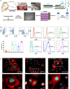
Fig. 1
Fig. 1
Isolation and characterization of human amniotic epithelial cells (hAECs). A Schematic representation of hAEC isolation protocol. Human amniotic membrane is harvested from term placenta, washed and hAECs are detached from the membrane using trypsin–EDTA. The resulting cells are then washed and cultured for 5–7 days. HAECs are then removed from the culture flasks at 80% confluence by mild trypsinization, and cryopreserved for later use. B hAECs were characterized by flow cytometry. Upper left panel shows the gating strategy used to obtain the quantification of positive populations for mesenchymal markers (CD105, CD90), pluripotency marker (SSEA-4), epithelial marker (CD326) and immunomodulatory markers (HLA-E and HLA-G). Representative examples are shown in the right panel, and quantification from 28 distinct hAEC batches (i.e. isolated from 28 placentae) is shown in the bottom left panel. C Immunofluorescent images of hAECs stained for the pluripotency markers SSEA-4 and Oct-4 and the immunomodulatory marker HLA-G (upper panels, scale bars = 100 um) and corresponding magnifications (lower panels, scale bars = 10 um)
Fig. 2
Fig. 2
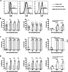
IFN-γ increases the expression of HLA-E and HLA-G in hAECs. hAEC monocultures were exposed to various concentration of IFN-γ for 24 and 48 h and were characterized by flow cytometry. A Representative flow cytometry histograms for CD105 (left panel), HLA-G (central panel) and HLA-E (right panel) positive populations in hAEC cultures exposed for 48 h to 1000 U/mL IFN-γ (dark lines) compared to untreated hAECs (grey lines). Isotype control is shown as dotted lines. Positive population for HLA-G (B), HLA-E (C) and CD105 (D), were quantified after exposition to low (left panels: 10, 25 and 50 U/mL, n = 4), medium (central panels: 100, 200 and 500 U/mL, n = 4) and high (right panels: 1000 and 2000 U/mL, n = 3) concentrations of IFN-γ. Grey bars: 24 h exposure, black bars: 48 h exposure. * p < 0.05, ** p < 0.01, **** p < 0.0001
Fig. 3
Fig. 3
hAECs secrete anti-inflammatory cytokines under pro-inflammatory conditions and protect rat islets against proinflammatory cytokine-induced damages in vitro. A Schematic representation of the experimental protocol. hAECs were cultured for 48 h before being exposed to a pro-inflammatory cytokines cocktail (100 U/mL IFN-γ, 800 U/mL TNF-α,50 u/mL IL-1β) for 48 h. Rat islets (RI) were added 24 h after hAEC seeding and were cultured on the hAEC monolayers for 24 h before cytokines exposure. Controls were untreated and cytokines-exposed RI monocultures. B IL6 (left, n = 6), IL10 (central, n = 4) and G-CSF (right, n = 6) secretion quantifications in the hAEC culture supernatants before, after 24 h and 48 h of cytokines cocktail exposure. C Expression changes in hAEC monocultures after cytokines cocktail exposure for anti-inflammatory cytokine genes (left), immunomodulatory genes (central) and IFN-γ signaling-related genes (right) (n = 4). D Apoptosis rate (i.e. relative quantification of cytoplasmic histone-associated DNA fragments) in RI and hAEC monocultures and in RI + hAEC co-cultures untreated or exposed to the cytokines cocktail for 48 h (n = 3). E Expression changes in rat islets cultured alone or with hAECs after cytokines cocktail exposure for the pro-apoptotic Nfkb1 and the anti-apoptotic Bcl2 genes (n = 4). F Islet function given as the secretion index during a glucose stimulated insulin secretion test performed on RI cultured alone or with hAECs in the presence or absence of pro-inflammatory cytokines (n = 3). Blue bars: Untreated cultures, red bars: cultures exposed to the cytokine cocktail. Empty bars: hAEC monocultures, patterned bars: RI monocultures, filled bars: RI + hAEC cocultures. * p < 0.05, ** p < 0.01, *** p < 0.001, **** p < 0.0001
Fig. 4
Fig. 4
Hypothetic mechanisms behind immunomodulation and cytoprotection conferred to pancreatic islet by hAECs. Interrupted lines: Inhibitory or stimulatory cytokine-induced cascades leading to the improved immunomodulatory and anti-inflammatory properties of hAECs under pro-inflammatory conditions. Bold lines: Signaling pathways leading to proinflammatory cytokine induced islet β cell apoptosis and loss of function reported in the literature. Dotted lines: Signaling pathways triggered by the anti-inflammatory and immunomodulatory factors secreted by hAECs. Red color indicates inhibitory pathways whereas green indicates activating pathways

References

    1. Arzouni AA, Vargas-Seymour A, Nardi N, King AJF, Jones PM. Using mesenchymal stromal cells in islet transplantation. Stem Cells Translational Medicine. 2018;7(8):559–563. doi: 10.1002/sctm.18-0033. - DOI - PMC - PubMed
    1. Banas RA, Trumpower C, Bentlejewski C, Marshall V, Sing G, Zeevi A. Immunogenicity and immunomodulatory effects of amnion-derived multipotent progenitor cells. Human Immunology. 2008;69(6):321–328. doi: 10.1016/j.humimm.2008.04.007. - DOI - PubMed
    1. Barshes NR, Wyllie S, Goss JA. Inflammation-mediated dysfunction and apoptosis in pancreatic islet transplantation: Implications for intrahepatic grafts. Journal of Leukocyte Biology. 2005;77(5):587–597. doi: 10.1189/jlb.1104649. - DOI - PubMed
    1. Carbone, A., Castellani, S., Favia, M., Diana, A., Paracchini, V., Di Gioia, S., … Conese, M. (2014). Correction of defective CFTR/ENaC function and tightness of cystic fibrosis airway epithelium by amniotic mesenchymal stromal (stem) cells. Journal of Cellular and Molecular Medicine,18(8), 1631–1643. 10.1111/jcmm.12303 - PMC - PubMed
    1. Cargnoni, A., Gibelli, L., Tosini, A., Signoroni, P. B., Nassuato, C., Arienti, D., … Parolini, O. (2009). Transplantation of allogeneic and xenogeneic placenta-derived cells reduces bleomycin-induced lung fibrosis. Cell Transplantation,18(4), 405–422. 10.3727/096368909788809857 - PubMed

Publication types