. 2021 Oct 6;13(614):eabe8868.
doi: 10.1126/scitranslmed.abe8868. Epub 2021 Oct 6.

Skeletal muscle regeneration with robotic actuation-mediated clearance of neutrophils

Affiliations

Skeletal muscle regeneration with robotic actuation-mediated clearance of neutrophils

Bo Ri Seo et al. Sci Transl Med. .

Abstract

Mechanical stimulation (mechanotherapy) can promote skeletal muscle repair, but a lack of reproducible protocols and mechanistic understanding of the relation between mechanical cues and tissue regeneration limit progress in this field. To address these gaps, we developed a robotic device equipped with real-time force control and compatible with ultrasound imaging for tissue strain analysis. We investigated the hypothesis that specific mechanical loading improves tissue repair by modulating inflammatory responses that regulate skeletal muscle regeneration. We report that cyclic compressive loading within a specific range of forces substantially improves functional recovery of severely injured muscle in mice. This improvement is attributable in part to rapid clearance of neutrophil populations and neutrophil-mediated factors, which otherwise may impede myogenesis. Insights from this work will help advance therapeutic strategies for tissue regeneration broadly.


Conflict of interest statement

Competing interests: D.J.M. and C.J.W. are inventors on patent/patent application (U.S. Patent Application no. 15/776,853, publication no. US 2020/0222582) held/submitted by Harvard University/Wyss Institute for this study. The authors declare that they have no other competing interests.

Figures

Fig. 1.
Fig. 1.. Electromagnetic linear actuator with integrated force sensor delivers reproducible compressive loading to muscle tissue.
(A) Photograph of robotic soft-interface actuator equipped with a force sensor and demonstration of the actuator positioned toward the injured tibialis anterior (TA) muscle of hindlimb of mouse. (B) Schematic illustration of the design and force-control mechanism of the actuator and setup with ultrasound transducer for real-time tissue monitoring. (C) Real-time monitoring of input and output loading force profiles (0.15, 0.3, and 0.6 N). (D) Representative ultrasound images of TA muscle captured during ML with 0.15, 0.3, and 0.6 N compressive forces and a scatter dot plot of corresponding tissue strains. (E) Representative strain heatmap of TA muscle obtained from computational simulation without or with ML (Ctrl versus 0.15 N). (n = 6 per condition, means ± SD). *P < 0.05, determined by Kruskal-Wallis test with post hoc Dunn’s tests.
Fig. 2.
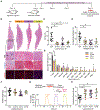
Fig. 2.. Cyclic loading improves skeletal muscle regeneration after severe injury.
(A) Experimental design and time line of TA muscle injury, ML treatment, and analysis in mice. (B) Representative hematoxylin and eosin (H&E) images of longitudinal histological sections of TA and its cross sections stained with H&E (top), Masson’s trichrome (collagen in blue, center), and laminin (bottom). Scale bars, 1 mm (entire TA) and 100 μm (cross sections). (C to G) Quantification of (C) fibrotic regions (appearing as blue in Masson’s trichome in B), (D) damaged muscle fibers (indicated by black arrows in H&E in B), (E) cross-sectional areas of muscle fibers, (F) muscle weight measured at the end point, (G) representative tetanic force graphs, and (H) measurement of normalized contraction force of TA muscle after 14-day treatment for control (no loading) and ML with various forces after the injury. Red dashed line indicates the contraction forces from uninjured TA muscle. Data in (C) to (G) are means ± SD (n = 4 to 16 per condition), and *P < 0.05, determined by Kruskal-Wallis test with post hoc Dunn’s tests.
Fig. 3.
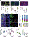
Fig. 3.. ML mediates rapid clearance of cytokines and neutrophils with effects on MPCs.
(A) Heatmap of a subset of cytokines in injured TA muscle with and without mechanical stimulation for 3 to 14 days (D3, D7, and D14). Data are represented as values in tissues treated with ML normalized to the untreated control tissue at the same time point. (B and C) Representative flow, dot plot, and immunofluorescence images on day 3 and quantification of neutrophil populations in muscle tissue without and with ML (0.3 N) for 14 days. Both pressure cuff and robotic actuator were used for mechanical stimulation. Scale bars, 500 and 50 μm, respectively (n = 4 to 12 per condition, means ± SD). *P < 0.05 determined by two-way ANOVA with Bonferroni’s multiple comparison tests and unpaired two-tailed Mann-Whitney test, respectively. (D) Immunofluorescence micrographs visualizing proliferation of MPCs (top) stained for EdU, MyHC, and DAPI and differentiation of MPCs (bottom) with desmin, MyHC, and DAPI staining after 3-or 5-day treatment with neutrophil-conditioned medium (+NeutCM) or control medium (−NeutCM), respectively. Scale bars, 100 μm. (E to G) Quantification of (E) EdU+ or MyHC+/desmin+ cell populations, (F) the population of multinucleated myotubes, and (G) distribution of myotube lengths, respectively. (n = 4 to 6 per condition, means ± SD). P < 0.05 (*) determined by unpaired two-tailed Mann-Whitney test. (H) Bar graphs of relative expression in log scale in NeutCM (relative to basal media) and TA muscle (ML-treated group versus control group). (I) Immunofluorescence micrographs visualizing proliferation of MPCs stained for EdU, MyHC, and DAPI and differentiation of MPCs with desmin, MyHC, and DAPI staining after 3-or 5-day treatment without and with NeutCM primed with neutralizing antibodies to different factors, and (J) quantification of EdU+ MPC populations and (K) desmin+/MyHC+ double positive myoblast populations (n = 6 per condition, means ± SD). Scale bars = 100 μm. *P < 0.05 determined by Kruskal-Wallis test with post hoc Dunn’s tests.
Fig. 4.
Fig. 4.. ML accelerates myogenesis and recovery of mature muscle fiber type composition.
(A to C) Representative immunofluorescence images and quantification of (A) MyoD+, (B) desmin+, and (C) Pax7+ MPCs in injured murine TA muscle after 3-day treatment with ML. Scale bars, 50 μm (n = 6 to 8 per condition, means ± SD), and *P < 0.05, determined by unpaired two-tailed Mann-Whitney test. (D and E) Immunofluorescence images and stacked bar graphs visualizing muscle fiber type compositions of injured TA muscle treated without and with ML for 14 days relative to uninjured TA muscle. Scale bars, 50 μm. (F to I) Quantification of muscle fiber subtypes of TA muscle: (F) type IIB (blue), (G) type IIX (black), (H) type IIA (green), and (I) type I (white). Magenta is laminin (n = 4 to 9 per condition, means ± SD), and *P < 0.05 determined by Kruskal-Wallis test with post hoc Dunn’s tests.
Fig. 5.
Fig. 5.. Antibody-mediated clearance of neutrophils mimics the effects of ML on muscle regeneration.
(A) Experimental design and time line of temporal depletion of neutrophils using treatment with anti-Ly6g antibody or ML on injured TA muscle in mice. (B) Representative H&E images of longitudinal and cross sections of TA muscle (top) treated without and with anti-Ly6g antibody or ML and associated tissue cross section stained with Masson’s trichrome (center) and stained for laminin (bottom). Scale bars, 1 mm and 100 μm, respectively. (C to F) Quantification of (C) fibrotic regions (interstitial blue regions in Masson’s trichrome in B), (D) damaged muscle fibers (H&E in B), and (E) measurement of CSAs of muscle fibers based on laminin-stained samples and (F) normalized contraction force of TA muscle 14 days after injury (n = 6 to 16 per condition, means ± SD), and *P < 0.05, determined by one-way ANOVA with post hoc Tukey’s tests.

References

    1. Liu J, Saul D, Böker KO, Ernst J, Lehman W, Schilling AF, Current methods for skeletal muscle tissue repair and regeneration. Biomed. Res. Int 2018, 1984879 (2018). - PMC - PubMed
    1. Turner NJ, Badylak SF, Regeneration of skeletal muscle. Cell Tissue Res. 347, 759–774 (2012). - PubMed
    1. Tidball JG, Regulation of muscle growth and regeneration by the immune system. Nat. Rev. Immunol 17, 165–178 (2017). - PMC - PubMed
    1. Kwee BJ, Seo BR, Najibi AJ, Li AW, Shih T-Y, White D, Mooney DJ, Treating ischemia via recruitment of antigen-specific T cells. Sci. Adv 5, eaav6313 (2019). - PMC - PubMed
    1. Raimondo TM, Mooney DJ, Functional muscle recovery with nanoparticle-directed M2 macrophage polarization in mice. Proc. Natl. Acad. Sci. U.S.A 115, 10648–10653 (2018). - PMC - PubMed

Publication types

LinkOut - more resources