. 2021 Dec;15(12):3596-3614.
doi: 10.1002/1878-0261.13110. Epub 2021 Oct 27.

Small extracellular vesicles from malignant ascites of patients with advanced ovarian cancer provide insights into the dynamics of the extracellular matrix

Affiliations

Small extracellular vesicles from malignant ascites of patients with advanced ovarian cancer provide insights into the dynamics of the extracellular matrix

Barbara Bortot et al. Mol Oncol. 2021 Dec.

Abstract

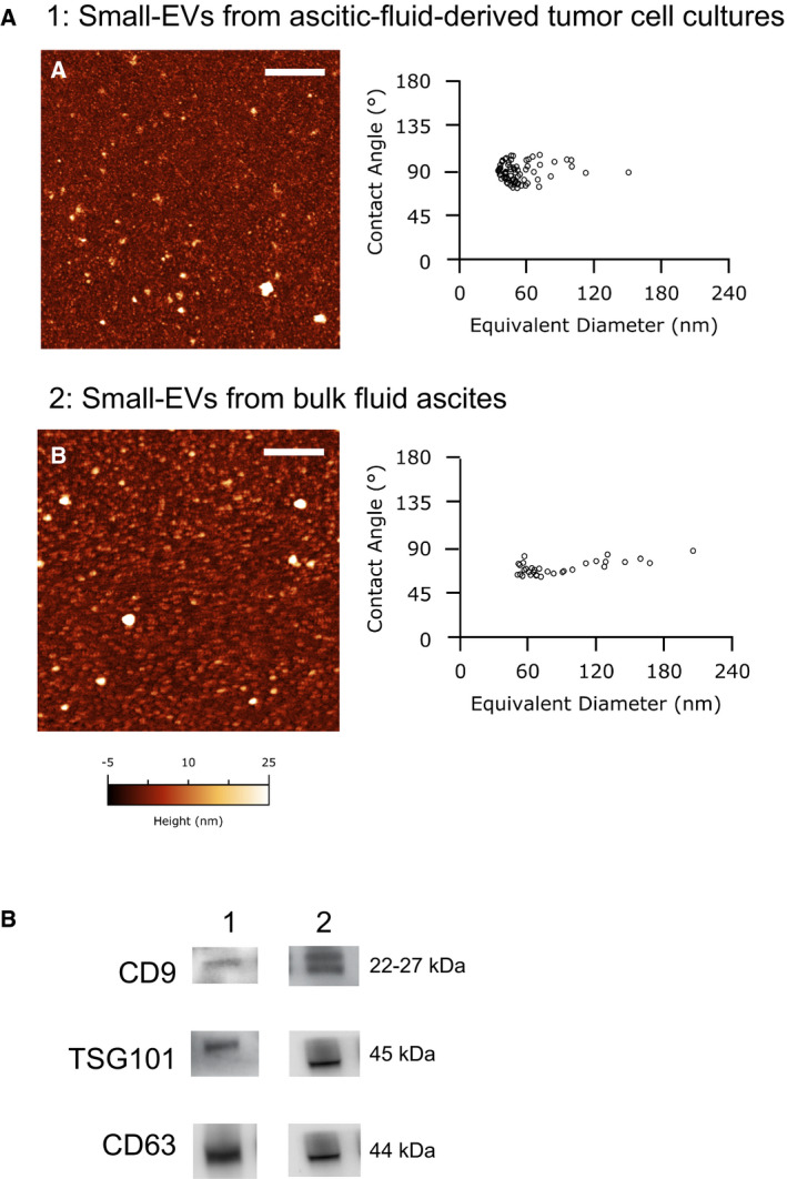
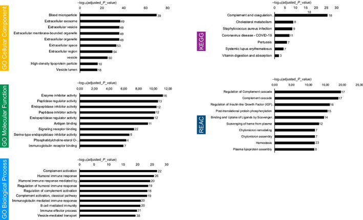
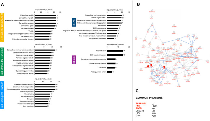
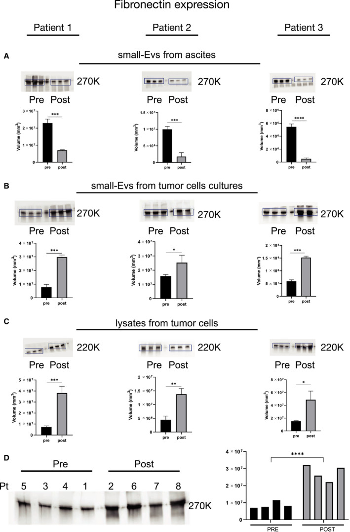
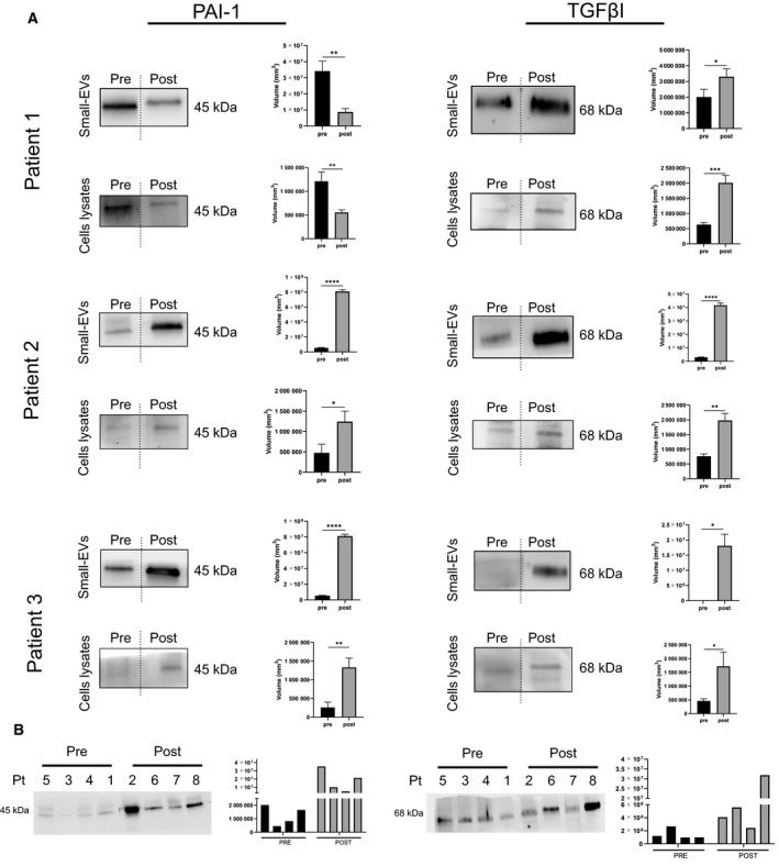
The exact role of malignant ascites in the development of intraperitoneal metastases remains unclear, and the mechanisms by which extracellular vesicles (EVs) promote tumor progression in the pre-metastatic niche have not been fully discovered. In this study, we characterized ascites from high-grade epithelial ovarian cancer patients. Small-EVs (30-150 nm) were isolated from two sources-the bulk ascites and the ascitic fluid-derived tumor cell cultures-and assessed with a combination of imaging, proteomic profiling, and protein expression analyses. In addition, Gene Ontology and pathway analysis were performed using different databases and bioinformatic tools. The results proved that the small-EVs derived from the two sources exhibited significantly different stiffness and size distributions. The bulk ascitic fluid-derived small-EVs were predominantly involved in the complement and coagulation cascade. Small-EVs derived from ascites cell cultures contained a robust proteomic profile of extracellular matrix remodeling regulators, and we observed an increase in transforming growth factor-β-I (TGFβI), plasminogen activator inhibitor 1 (PAI-1), and fibronectin expression after neoadjuvant chemotherapy. When measured in the two sources, we demonstrated that fibronectin exhibited opposite expression patterns in small-EVs in response to chemotherapy. These findings highlight the importance of an ascites cell isolation workflow in investigating the treatment-induced cancer adaption processes.

Keywords: ascites; chemotherapy; extracellular matrix; extracellular vesicles; fibronectin; ovarian cancer.


Conflict of interest statement

The authors declare no conflict of interest.

Figures

Fig. 1
Fig. 1
CA125 kinetic and systemic inflammatory response markers. Measurement of different parameters which are considered independent predictors of chemotherapy response. (A) The graph reports changes in CA125 serum levels in the patient cohort throughout chemotherapy. U·mL−1: units per milliliters. (B) The table reports values in CA125 serum levels, and their percentage decrease between T0 and T1. T0: time of diagnosis before starting neoadjuvant chemotherapy (NACT). T1: time of interval debulking surgery (IDS). (C) PLR: platelet‐to‐lymphocyte ratio. (D) NLR: neutrophil‐to‐lymphocyte ratio. The range considered normal for PLR (60.0–239.0) and NLR (0.9–3.9) is highlighted in red.
Fig. 2
Fig. 2
Morphology and identification of ascites‐derived epithelial ovarian cancer (EOC) cells. (A) Representative phase‐contrast images of EOC cells. In the upper image: cells after initial isolation, grape‐like cluster (arrow), and single EOC cells. In the lower image: a confluent monolayer of EOC cells expanded in culture. Note the typical epithelial cobblestone morphology and tight cell‐to‐cell junction. (B) Subsequent cytological analysis. Immunochemistry of EOC cells showed expression of epithelial marker multicytokeratin, mesenchymal marker, vimentin, and cell proliferation marker Ki67. (C) Immunofluorescence staining of EOC cells revealed the expression of the Mucin‐16 tumor marker (CA125). Brightfield, nucleus staining with blu DAPI, Mucin‐16 marker staining with green Alexa Fluor® 488, and merge image (DAPI+ Alexa Fluor® 488) are reported. Bar = 100 μm. For each set of analyses, representative results out of three independent experiments are shown.
Fig. 3
Fig. 3
Characterization of small‐EVs isolated from human ovarian ascites fluid. (A) Representative liquid Atomic Force Microscopy (AFM) micrographs of small‐EVs from ascites‐derived tumor cells (A) and small‐EVs from bulk fluid ascites (B), the length of both scale bars is 1 µm. On the right: Contact Angle vs Diameter scatterplot of small‐EVs. Each circle represents one individual exosome as measured via AFM imaging in liquid. (B) CD63, CD9, and TSG101 were detected by western blot in small‐EVs extracted from ascites‐derived tumor cell cultures (1) and bulk fluid ascites small‐EVs (2). For each set of analyses, representative results out of three independent experiments are shown.
Fig. 4
Fig. 4
Proteomic profiling of the small‐EVs from bulk fluid ascites. Gene lists derived from proteomics data (n = 8) underwent KEGG, Gene Ontology (GO), and Reactome (REAC) enrichment analysis using g: Profiler. Negative log10 of adjusted P‐value and intersections size are reported in graphs.
Fig. 5
Fig. 5
Proteomic profiling of the small‐EVs from ascites‐derived tumor cells. (A) Gene lists derived from proteomics data (n = 8) underwent KEGG, Gene Ontology (GO), and Reactome (REAC) enrichment analysis using g:Profiler. Negative log10 of adjusted P‐value and intersections size are reported in graphs. (B) Protein–protein interaction network visualized by BioDiscoverTM. Interactions with supporting evidence from pathway resources are highlighted in blue. Fibronectin, TGFβI, and PAI‐1 are highlighted in red. (C) 12 proteins were identified that were common to every EV sample.
Fig. 6
Fig. 6
Differential fibronectin expression in small‐EV samples taken before and after chemotherapy. Protein extracts (30 µg) prepared from small‐EVs isolated from bulk fluid ascites (A), small‐EVs isolated from ascites tumor cell cultures (B), and ascites tumor cells lysates (C) of patients 1, 2, and 3 were loaded on a 4–12% SDS/PAGE followed by western blot using antibodies against fibronectin. Two‐tailed t‐tests were used for protein expression analysis. Data are reported as means ± SD of one experiment performed in triplicate. *P < 0.05, **P < 0.01, ***P < 0.001, ****P < 0.0001. (D) Analysis of the small‐EVs isolated from ascites tumor cell cultures on the cohort of patients divided into two groups: the patients who did not receive chemotherapy at the time of sampling and the patients who underwent chemotherapy. ****P < 0.0001. The patient’s number corresponds to Table 1.
Fig. 7
Fig. 7
Differential TGFβI and PAI‐1 expression in small‐EV samples taken before and after chemotherapy. (A) Protein extracts (30 µg) prepared from small‐EVs isolated from bulk fluid ascites, small‐EVs isolated from ascites tumor cell cultures, and ascites tumor cell lysates of patients 1, 2, and 3 were loaded on a 4–12% SDS/PAGE followed by western blot using antibodies against TGFβI and PAI‐1. Two‐tailed t‐tests were used for protein expression analysis. Data are reported as means ± SD of one experiment performed in triplicate. *P < 0.05, **P < 0.01, ***P < 0.001, ****P < 0.0001. (B) Analysis of the small‐EVs isolated from ascites tumor cell cultures on the cohort of patients divided into two groups: the patients who did not receive chemotherapy at the time of sampling and the patients who underwent chemotherapy. The patient’s number corresponds to Table 1.

References

    1. Henderson JT, Webber EM & Sawaya GF (2018) Screening for ovarian cancer: updated evidence report and systematic review for the US Preventive Services Task Force. JAMA 319, 595–606. - PubMed
    1. Di Lorenzo G, Ricci G, Severini GM, Romano F & Biffi S (2018) Imaging and therapy of ovarian cancer: clinical application of nanoparticles and future perspectives. Theranostics 8, 4279–4294. - PMC - PubMed
    1. Jayson GC, Kohn EC, Kitchener HC & Ledermann JA (2014) Ovarian cancer. Lancet Lond Engl 384, 1376–1388. - PubMed
    1. Agarwal R & Kaye SB (2003) Ovarian cancer: strategies for overcoming resistance to chemotherapy. Nat Rev Cancer 3, 502–516. - PubMed
    1. Bogani G, Rossetti D, Ditto A, Martinelli F, Chiappa V, Mosca L, Leone Roberti Maggiore U, Ferla S, Lorusso D & Raspagliesi F (2018) Artificial intelligence weights the importance of factors predicting complete cytoreduction at secondary cytoreductive surgery for recurrent ovarian cancer. J Gynecol Oncol 29, e66. - PMC - PubMed

Publication types