Review
. 2022 Feb 10:84:435-459.
doi: 10.1146/annurev-physiol-060821-083306. Epub 2021 Oct 6.

Mitochondria and Inflammatory Bowel Diseases: Toward a Stratified Therapeutic Intervention

Affiliations
Review

Mitochondria and Inflammatory Bowel Diseases: Toward a Stratified Therapeutic Intervention

Gwo-Tzer Ho et al. Annu Rev Physiol. .

Abstract

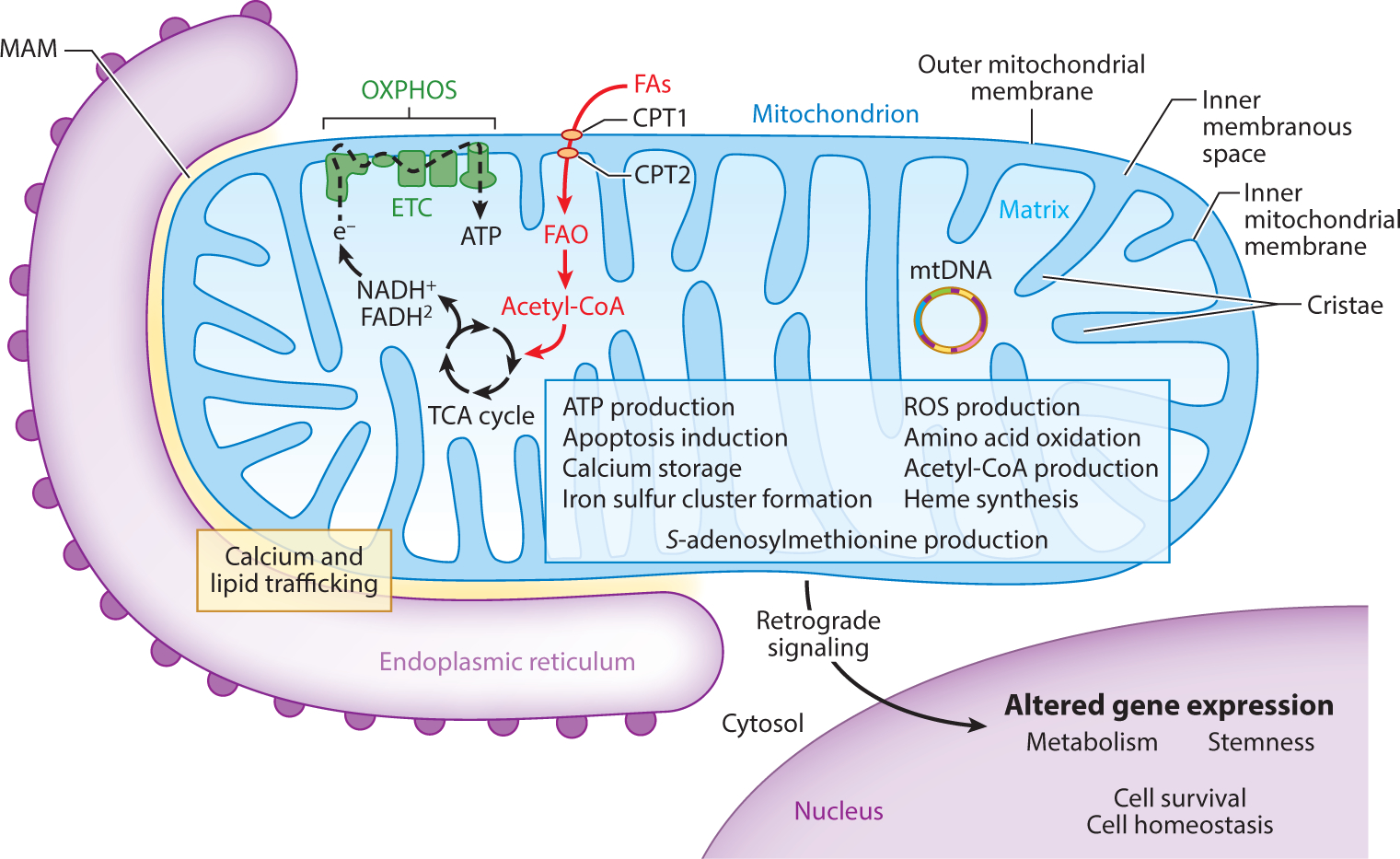
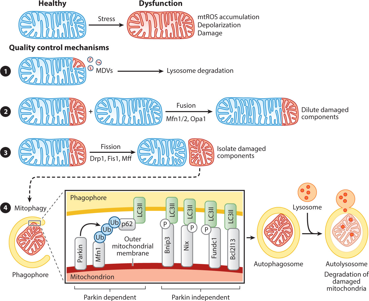
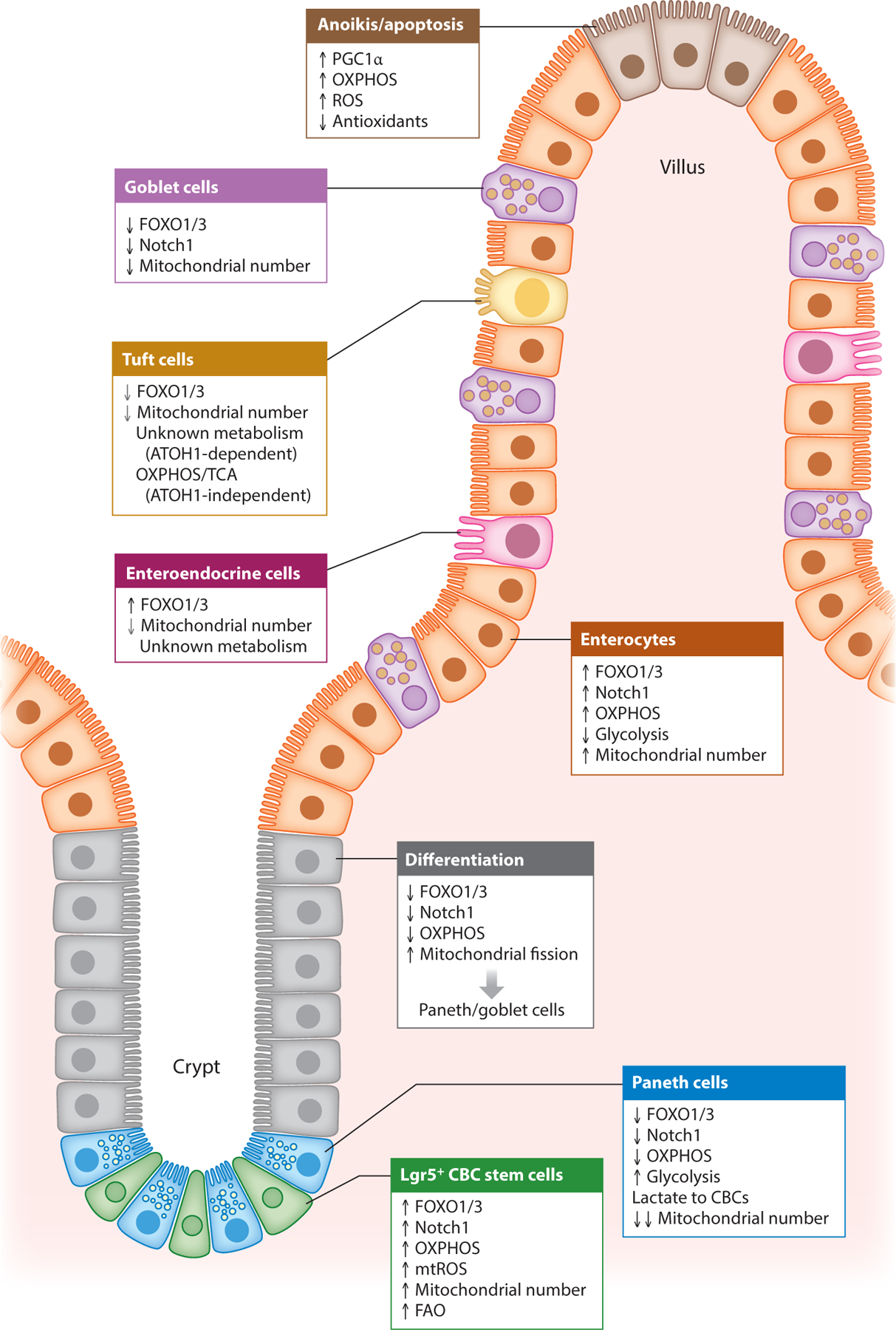
Mitochondria serve numerous critical cellular functions, rapidly responding to extracellular stimuli and cellular demands while dynamically communicating with other organelles. Mitochondrial function in the gastrointestinal epithelium plays a critical role in maintaining intestinal health. Emerging studies implicate the involvement of mitochondrial dysfunction in inflammatory bowel disease (IBD). This review presents mitochondrial metabolism, function, and quality control that converge in intestinal epithelial stemness, differentiation programs, barrier integrity, and innate immunity to influence intestinal inflammation. Intestinal and disease characteristics that set the stage for mitochondrial dysfunction being a key factor in IBD and, in turn, pathogenic mitochondrial mechanisms influencing and potentiating the development of IBD, are discussed. These findings establish the basis for potential mitochondrial-targeted interventions for IBD therapy.

Keywords: Crohn's disease; inflammation; intestinal epithelium; mitochondria; ulcerative colitis.


Figures

Figure 1
Figure 1
Mitochondrial structure is directly related to function. The structure of mitochondria facilitates numerous functions based on cellular need (blue box) (–, , –12). Arguably, the most important function performed by mitochondria is energy production in the form of ATP through OXPHOS. In Lgr5+ crypt base columnar stem cells, FAO is emerging as an important provider of acetyl-CoA to fuel the TCA cycle and OXPHOS (–39). Mitochondria also actively communicate with other organelles important for global cell homeostasis. Mitochondria are intimately associated with the endoplasmic reticulum through MAMs, where the exchange of lipids and calcium influences homeostasis of each organelle (11). Retrograde signaling serves as mitochondrial-to-nuclear intraorganelle communication, resulting in altered gene expression based on mitochondrial status (12). Abbreviations: Acetyl-CoA, acetyl-coenzyme A; ATP, adenosine triphosphate; CPT, carnitine palmitoyltransferase; e, donated electron; ETC, electron transport chain; FAs, fatty acids; FAO, fatty acid oxidation; MAM, mitochondrial-associated membrane; mtDNA, mitochondrial DNA; OXPHOS, oxidative phosphorylation; ROS, reactive oxygen species; TCA, tricarboxylic acid.
Figure 2 :
Figure 2 :
Mitochondrial quality control mechanisms. Mitochondrial dysfunction can be controlled by various mechanisms. (❶) The release of MDVs containing damaged mitochondrial proteins destined for degradation by the lysosomes is considered a first round of defense for the organelle (13). The processes of mitochondrial fission and fusion are mediated by GTPases of the dynamin family. (❷) Mitochondrial fusion, mediated by Mfn1, Mfn2, and Opa1, facilitates the sharing of mtDNA between mitochondria, thereby providing more support for critical functions such as oxidative phosphorylation (14). (❸) Mitochondrial fission, mediated by Drp1, Fis1, and Mff, isolates damaged from healthy components and is crucial for mitophagy by dividing mitochondria into small enough sizes to fit into the autophagosome. (❹) Damaged mitochondria are marked for mitophagy by OMM-localized autophagy receptors, which are either dependent on the E3 ubiquitin ligase Parkin or are Parkin independent. Parkin is recruited to damaged mitochondria by Pink1 and forms polyubiquitinated chains on OMM proteins (such as Mfn1, Mfn2, or VDAC1) that are then recognized by an autophagy receptor (such as p62/sequestosome 1, optineurin, NBR1, or AMBRA1). These autophagy receptors bind to LC3II present in the membrane of the forming phagosome, thereby incorporating the damaged mitochondrion into the autophagy pathway for ultimate degradation in the autolysosome. Parkin-independent autophagy receptors contain transmembrane domains and upon expression constitutively localize to the OMM and interact with autophagy factors such as LC3II (via an LIR domain), ULK1, DFCP1, and WIPI1. Expression of Bnip3 and Nix are controlled by the transcription factors Hif1α, NF-κB, or FOXO3, suggesting that hypoxia, inflammation, and stemness pathways are involved in mitophagy induction (142, 143). Posttranslational modification of Bnip3, Nix, and Fundc1 by phosphorylation within the LIR domain increases their affinity for LC3II binding, suggesting that phosphorylation regulates their pro-mitophagy activity (144). Abbreviations: AMBRA1, autophagy and beclin 1 regulator 1; DFCP1, double FYVE-containing protein 1; Drp1, dynamin-related protein 1; Fis1, fission 1; FOXO3, Forkhead box O 3; GTPases, guanosine triphosphatases; Hif1α, hypoxia-inducible factor 1 alpha; LC3, microtubule-associated protein 1A/1B-light chain 3; LIR, LC3 interacting region; MDV, mitochondrial-derived vesicle; Mff, mitochondria fission factor; Mfn, mitofusin; mtDNA, mitochondrial DNA; mtROS, mitochondrial-derived reactive oxygen species; NBR1, next to BRCA1 gene 1; NF-κB, nuclear factor kappa B; OMM, outer mitochondrial membrane; Opa1, optic atrophy 1; P, phosphorylation; Ub, ubiquitin; ULK1, Unc-51 like autophagy activating kinase 1; VDAC1, voltage-dependent anion channel 1; WIPI1, WD repeat domain phosphoinositide interacting 1.
Figure 3 :
Figure 3 :
Mitochondrial influence on intestinal epithelial cell fate. Mitochondrial health, metabolism, and numbers play important roles in driving CBC stemness and determining the differentiation of secretory versus absorptive epithelial lineages. CBCs exhibit high mitochondrial respiration and numbers dependent on high expression of FOXO1/3 (27, 28). During differentiation to secretory cells (Paneth cells), a metabolic switch away from OXPHOS is driven by inhibition of FOXO1/3 and enhanced mitochondrial fission (27). During differentiation to absorptive enterocytes, dependency on OXPHOS and expression of FOXO1/3 remains unchanged. Cell death of apical IECs furthest from the crypt base is also influenced by mitochondrial metabolism driven by PGC1α expression (41). Grey arrows indicate that the observed effect is to a lesser extent. Abbreviations: CBC, Lgr5+ crypt base columnar stem cell; IEC, intestinal epithelial cell; FAO, fatty acid oxidation; FOXO, Forkhead box O; mtROS, mitochondrial-derived ROS; OXPHOS, oxidative phosphorylation; PGC1α, peroxisome proliferator-activated receptor-γ coactivator 1-α; ROS, reactive oxygen species; TCA, tricarboxylic acid.

References

    1. Ni HM, Williams JA, Ding WX. 2015. Mitochondrial dynamics and mitochondrial quality control. Redox Biol. 4:6–13 - PMC - PubMed
    1. Frey TG, Mannella CA. 2000. The internal structure of mitochondria. Trends Biochem. Sci. 25:319–24 - PubMed
    1. Ryan MT, Hoogenraad NJ. 2007. Mitochondrial-nuclear communications. Annu. Rev. Biochem. 76:701–22 - PubMed
    1. Kuhlbrandt W 2015. Structure and function of mitochondrial membrane protein complexes. BMC Biol. 13:89. - PMC - PubMed
    1. Minocherhomji S, Tollefsbol TO, Singh KK. 2012. Mitochondrial regulation of epigenetics and its role in human diseases. Epigenetics 7:326–34 - PMC - PubMed

Publication types

MeSH terms

LinkOut - more resources