. 2021 Oct 12;37(2):109825.
doi: 10.1016/j.celrep.2021.109825. Epub 2021 Sep 28.

B.1.617.2 enters and fuses lung cells with increased efficiency and evades antibodies induced by infection and vaccination

Affiliations

B.1.617.2 enters and fuses lung cells with increased efficiency and evades antibodies induced by infection and vaccination

Prerna Arora et al. Cell Rep. .

Abstract

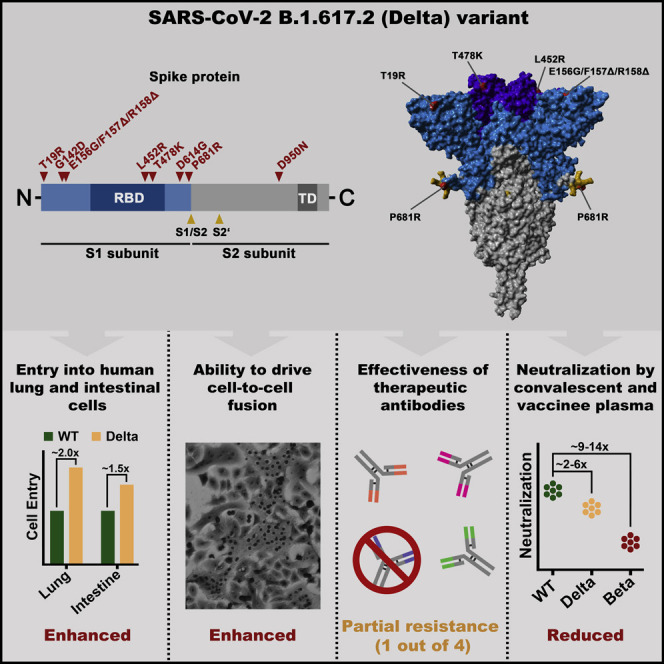
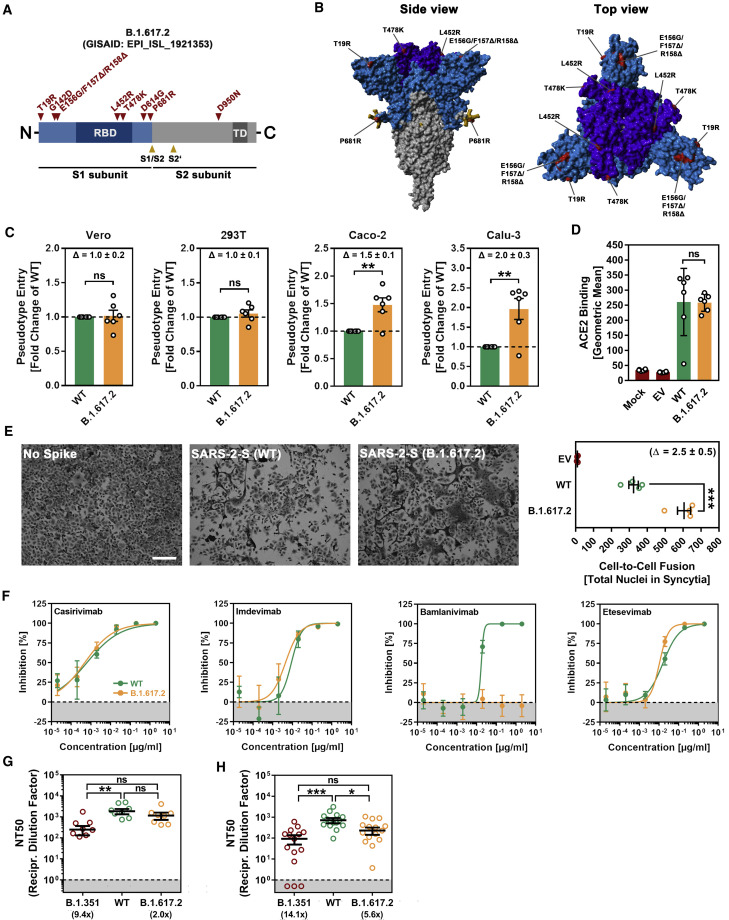
The Delta variant of severe acute respiratory syndrome coronavirus 2 (SARS-CoV-2), B.1.617.2, emerged in India and has spread to over 80 countries. B.1.617.2 replaced B.1.1.7 as the dominant virus in the United Kingdom, resulting in a steep increase in new infections, and a similar development is expected for other countries. Effective countermeasures require information on susceptibility of B.1.617.2 to control by antibodies elicited by vaccines and used for coronavirus disease 2019 (COVID-19) therapy. We show, using pseudotyping, that B.1.617.2 evades control by antibodies induced upon infection and BNT162b2 vaccination, although to a lesser extent as compared to B.1.351. We find that B.1.617.2 is resistant against bamlanivimab, a monoclonal antibody with emergency use authorization for COVID-19 therapy. Finally, we show increased Calu-3 lung cell entry and enhanced cell-to-cell fusion of B.1.617.2, which may contribute to augmented transmissibility and pathogenicity of this variant. These results identify B.1.617.2 as an immune evasion variant with increased capacity to enter and fuse lung cells.

Keywords: B.1.617.2; COVID-19; SARS-CoV-2; antibody; neutralization; spike.


Conflict of interest statement

Declaration of interests The authors declare no competing interests.

Figures

None
Graphical abstract
Figure 1
Figure 1
The spike protein of SARS-CoV-2 B.1.617.2 promotes efficient entry into human lung and colon cells, causes more cell-to-cell fusion, and evades antibody-mediated neutralization (A) Schematic overview of the S protein from SARS-CoV-2 variant B.1.617.2. RBD, receptor-binding domain; TD, transmembrane domain. (B) Location of the mutations found in SARS-CoV-2 variant B.1.617.2 in the context of the trimeric spike protein (color code: light blue, S1 subunit with RBD in dark blue; gray, S2 subunit; orange, S1/S2 and S2′ cleavage sites; red, mutated amino acid residues). (C) Pseudotyped particles bearing the S protein of wild-type (WT) SARS-CoV-2 or variant B.1.617.2 were inoculated onto the indicated cell lines, and transduction efficiency was quantified by measuring virus-encoded luciferase activity in cell lysates at 16 to 18 h post transduction. Presented are the average (mean) data from 6 biological replicates (each conducted with technical quadruplicates) for which transduction was normalized against SARS-CoV-2 S WT (= 1). Error bars indicate the standard error of the mean (SEM). Statistical significance of differences between WT and B.1.617.2 S proteins was analyzed by two-tailed Student’s t test (p > 0.05, not significant [ns]; ∗∗p ≤ 0.01). See also Figure S1A. (D) BHK-21 expressing the S protein of WT SARS-CoV-2 or variant B.1.617.2 were subsequently incubated with soluble ACE2 (harboring a C-terminal Fc-tag derived from human IgG) and AlexaFluor-488-conjugated anti-human antibody before being subjected to flow cytometry. ACE2 binding efficiency was analyzed by measuring the geometric mean channel fluorescence at 488 nm. Untransfected cells and cells transfected with empty expression plasmid served as controls. Presented are the average (mean) data from 6 biological replicates (each conducted with single samples). Error bars indicate the standard deviation (SD). Statistical significance of differences between WT and variant B.1.617.2 S proteins was analyzed by two-tailed Student’s t test (p > 0.05, ns). (E) Analysis of S protein-induced cell-to-cell fusion. A549-ACE2 cells were transfected with expression plasmid for the indicated S proteins or empty vector (EV). At 24 h post transfection, cells were fixed and subsequently stained with May-Gruenwald and Giemsa solutions. Presented are representative microscopic images (scale bar, 200 μm). For quantification of fusion efficiency, the total number of nuclei in syncytia per image was counted. Presented are the average (mean) data from 4 biological replicates (each conducted with single samples; for each sample, 3 randomly selected areas were imaged and independently analyzed by 2 persons). Error bars indicate the SEM. Statistical significance of differences between WT and B.1.617.2 S proteins was analyzed by two-tailed Student’s t test (∗∗∗p ≤ 0.001). (F) Neutralization of particles bearing SARS-CoV-2 WT or B.1.617.2 S proteins by monoclonal antibodies used for COVID-19 therapy. Pseudotyped particles bearing the S protein of WT SARS-CoV-2 or variant B.1.617.2 were incubated for 30 min at 37°C in the presence of escalating concentrations (0.00002, 0.0002, 0.002, 0.02, 0.2, and 2 μg/mL) of the indicated SARS-CoV-2 S protein-specific monoclonal antibody (please see Figure S1B) or an unrelated control antibody (please see Figure S1C) before being inoculated onto Vero cells. Transduction efficiency was quantified by measuring virus-encoded luciferase activity in cell lysates at 16 to 18 h post transduction. Presented are the average (mean) data from a single biological replicate (conducted with technical quadruplicates) for which transduction was normalized against samples that did not contain any antibody (= 0% inhibition). Error bars indicate the SD. The results were confirmed in a separate experiment. (G) Neutralization of particles bearing SARS-CoV-2 WT, B.1.351, or B.1.617.2 S proteins by antibodies in convalescent plasma. Pseudotyped particles bearing the S protein of SARS-CoV-2 WT, B.1.351 or B.1.617.2 were incubated for 30 min at 37°C in the presence of different dilutions of convalescent plasma (1:25, 1:100, 1:400, 1:1,600, 1:6,400, and 1:25,600). Transduction efficiency was quantified by measuring virus-encoded luciferase activity in cell lysates at 16 to 18 h post transduction and used to calculate the plasma dilution factor that leads to 50% reduction in S protein-driven cell entry (neutralizing titer 50 [NT50]). Presented are the data from a single biological replicate (conducted with technical quadruplicates) for a total of 8 convalescent plasma (black lines indicate the median; error bars indicate the SEM). Statistical significance of differences between the indicated groups was analyzed by Kruskal-Wallis analysis with Dunn’s post hoc test (p > 0.05, ns; p ≤ 0.05; ∗∗p ≤ 0.01; ∗∗∗p ≤ 0.001). Please see also Figure S1D. (H) The experiment was performed as described for (G), but this time serum from Comirnaty/BNT162b2-vaccinated individuals was investigated. Presented are the data from a single biological replicate (conducted with technical quadruplicates) for a total of 15 vaccinee sera (black lines indicate the median; error bars indicate the SEM). Please see also Figure S1E.

References

    1. Barnes C.O., West A.P., Jr., Huey-Tubman K.E., Hoffmann M.A.G., Sharaf N.G., Hoffman P.R., Koranda N., Gristick H.B., Gaebler C., Muecksch F., et al. Structures of Human Antibodies Bound to SARS-CoV-2 Spike Reveal Common Epitopes and Recurrent Features of Antibodies. Cell. 2020;182:828–842.e16. - PMC - PubMed
    1. Berger Rentsch M., Zimmer G. A vesicular stomatitis virus replicon-based bioassay for the rapid and sensitive determination of multi-species type I interferon. PLoS ONE. 2011;6:e25858. - PMC - PubMed
    1. Bussani R., Schneider E., Zentilin L., Collesi C., Ali H., Braga L., Volpe M.C., Colliva A., Zanconati F., Berlot G., et al. Persistence of viral RNA, pneumocyte syncytia and thrombosis are hallmarks of advanced COVID-19 pathology. EBioMedicine. 2020;61:103104. - PMC - PubMed
    1. Cai Y., Zhang J., Xiao T., Peng H., Sterling S.M., Walsh R.M., Jr., Rawson S., Rits-Volloch S., Chen B. Distinct conformational states of SARS-CoV-2 spike protein. Science. 2020;369:1586–1592. - PMC - PubMed
    1. Campbell F., Archer B., Laurenson-Schafer H., Jinnai Y., Konings F., Batra N., Pavlin B., Vandemaele K., Van Kerkhove M.D., Jombart T., et al. Increased transmissibility and global spread of SARS-CoV-2 variants of concern as at June 2021. Euro Surveill. 2021;26:2100509. - PMC - PubMed

Publication types

MeSH terms