MRI and CT coronary angiography in survivors of COVID-19
- PMID: 34615668
- PMCID: PMC8503921
- DOI: 10.1136/heartjnl-2021-319926
MRI and CT coronary angiography in survivors of COVID-19
Abstract
Objectives: To determine the contribution of comorbidities on the reported widespread myocardial abnormalities in patients with recent COVID-19.
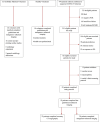
Methods: In a prospective two-centre observational study, patients hospitalised with confirmed COVID-19 underwent gadolinium and manganese-enhanced MRI and CT coronary angiography (CTCA). They were compared with healthy and comorbidity-matched volunteers after blinded analysis.
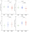
Results: In 52 patients (median age: 54 (IQR 51-57) years, 39 males) who recovered from COVID-19, one-third (n=15, 29%) were admitted to intensive care and a fifth (n=11, 21%) were ventilated. Twenty-three patients underwent CTCA, with one-third having underlying coronary artery disease (n=8, 35%). Compared with younger healthy volunteers (n=10), patients demonstrated reduced left (ejection fraction (EF): 57.4±11.1 (95% CI 54.0 to 60.1) versus 66.3±5 (95 CI 62.4 to 69.8)%; p=0.02) and right (EF: 51.7±9.1 (95% CI 53.9 to 60.1) vs 60.5±4.9 (95% CI 57.1 to 63.2)%; p≤0.0001) ventricular systolic function with elevated native T1 values (1225±46 (95% CI 1205 to 1240) vs 1197±30 (95% CI 1178 to 1216) ms;p=0.04) and extracellular volume fraction (ECV) (31±4 (95% CI 29.6 to 32.1) vs 24±3 (95% CI 22.4 to 26.4)%; p<0.0003) but reduced myocardial manganese uptake (6.9±0.9 (95% CI 6.5 to 7.3) vs 7.9±1.2 (95% CI 7.4 to 8.5) mL/100 g/min; p=0.01). Compared with comorbidity-matched volunteers (n=26), patients had preserved left ventricular function but reduced right ventricular systolic function (EF: 51.7±9.1 (95% CI 53.9 to 60.1) vs 59.3±4.9 (95% CI 51.0 to 66.5)%; p=0.0005) with comparable native T1 values (1225±46 (95% CI 1205 to 1240) vs 1227±51 (95% CI 1208 to 1246) ms; p=0.99), ECV (31±4 (95% CI 29.6 to 32.1) vs 29±5 (95% CI 27.0 to 31.2)%; p=0.35), presence of late gadolinium enhancement and manganese uptake. These findings remained irrespective of COVID-19 disease severity, presence of myocardial injury or ongoing symptoms.
Conclusions: Patients demonstrate right but not left ventricular dysfunction. Previous reports of left ventricular myocardial abnormalities following COVID-19 may reflect pre-existing comorbidities.
Trial registration number: NCT04625075.
Keywords: COVID-19; magnetic resonance imaging.
© Author(s) (or their employer(s)) 2022. Re-use permitted under CC BY-NC. No commercial re-use. See rights and permissions. Published by BMJ.
Conflict of interest statement
Competing interests: None declared.
Figures


Comment in
-
Cardiac involvement in COVID-19: cause or consequence of severe manifestations?Heart. 2022 Jan;108(1):7-8. doi: 10.1136/heartjnl-2021-320246. Epub 2021 Oct 16. Heart. 2022. PMID: 34656975 No abstract available.
References
Publication types
MeSH terms
Substances
Associated data
Grants and funding
- CH/11/2/28733/BHF_/British Heart Foundation/United Kingdom
- CS/17/1/32445/BHF_/British Heart Foundation/United Kingdom
- FS/17/79/33226/BHF_/British Heart Foundation/United Kingdom
- FS/TF/21/33008/BHF_/British Heart Foundation/United Kingdom
- RE/18/5/34216/BHF_/British Heart Foundation/United Kingdom
- FS/ICRF/20/26002/BHF_/British Heart Foundation/United Kingdom
- MR/T029153/1/MRC_/Medical Research Council/United Kingdom
- FS/14/78/31020/BHF_/British Heart Foundation/United Kingdom
- RG/16/10/32375/BHF_/British Heart Foundation/United Kingdom
- CH/09/002/BHF_/British Heart Foundation/United Kingdom
- PCL/17/04/CSO_/Chief Scientist Office/United Kingdom
- WT_/Wellcome Trust/United Kingdom
- CH/09/002/26360/BHF_/British Heart Foundation/United Kingdom
LinkOut - more resources
Full Text Sources
Medical
