. 2021 Oct 12;118(41):e2100198118.
doi: 10.1073/pnas.2100198118.

Structural basis of rotavirus RNA chaperone displacement and RNA annealing

Affiliations

Structural basis of rotavirus RNA chaperone displacement and RNA annealing

Jack P K Bravo et al. Proc Natl Acad Sci U S A. .

Abstract

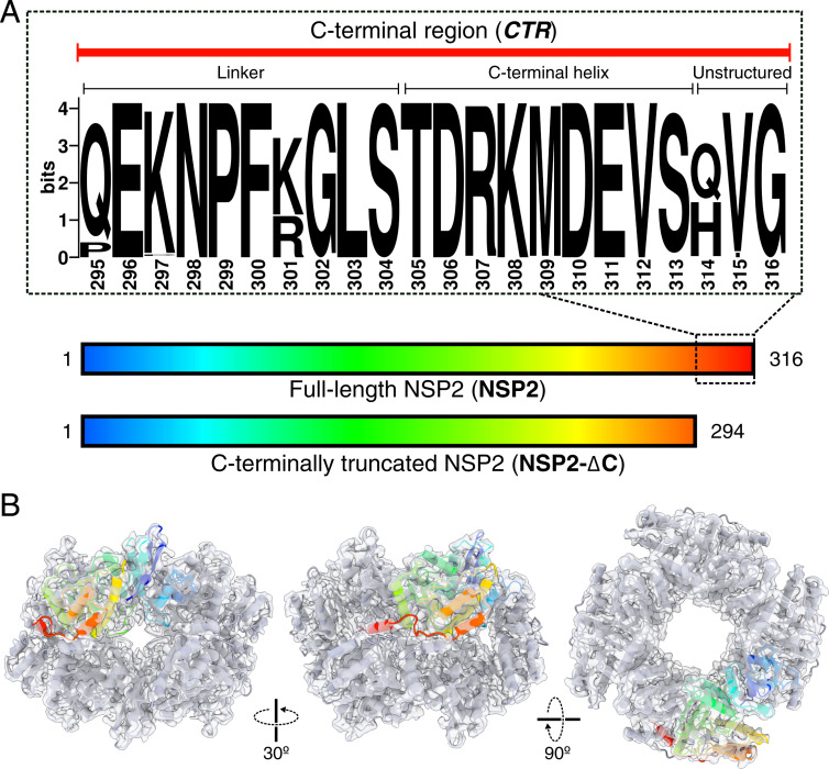
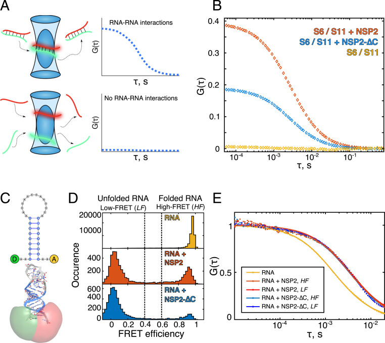
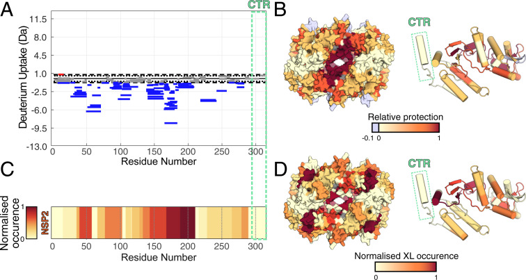
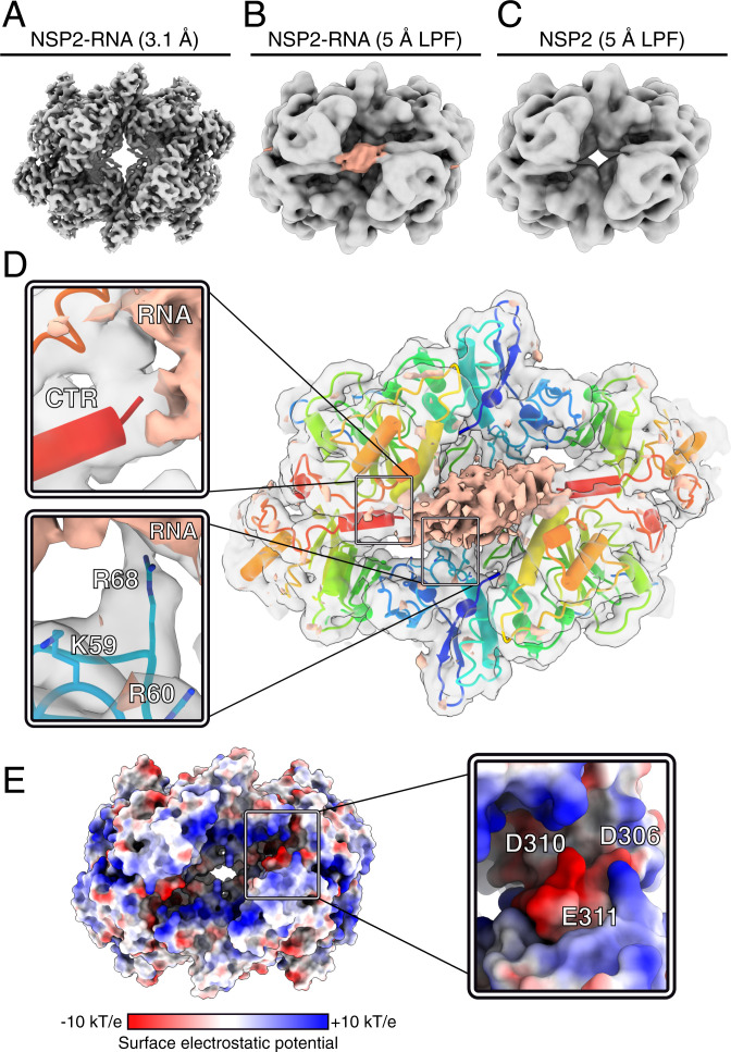
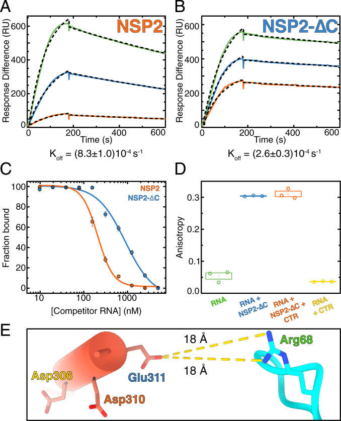
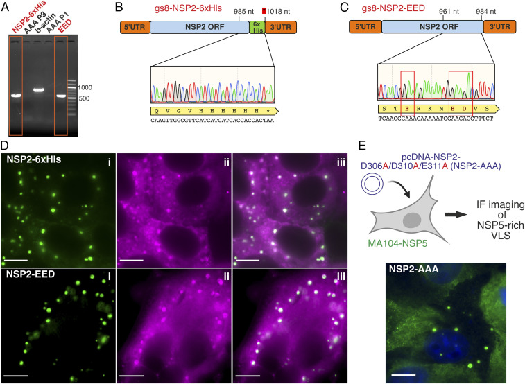
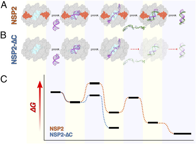
Rotavirus genomes are distributed between 11 distinct RNA molecules, all of which must be selectively copackaged during virus assembly. This likely occurs through sequence-specific RNA interactions facilitated by the RNA chaperone NSP2. Here, we report that NSP2 autoregulates its chaperone activity through its C-terminal region (CTR) that promotes RNA-RNA interactions by limiting its helix-unwinding activity. Unexpectedly, structural proteomics data revealed that the CTR does not directly interact with RNA, while accelerating RNA release from NSP2. Cryo-electron microscopy reconstructions of an NSP2-RNA complex reveal a highly conserved acidic patch on the CTR, which is poised toward the bound RNA. Virus replication was abrogated by charge-disrupting mutations within the acidic patch but completely restored by charge-preserving mutations. Mechanistic similarities between NSP2 and the unrelated bacterial RNA chaperone Hfq suggest that accelerating RNA dissociation while promoting intermolecular RNA interactions may be a widespread strategy of RNA chaperone recycling.

Keywords: RNA chaperones; genome assembly; ribonucleoproteins; rotavirus.


Conflict of interest statement

The authors declare no competing interest.

Figures

Fig. 1.
Fig. 1.
Structure and conservation of NSP2 CTR. (A) Constructs of full-length NSP2 (NSP2) and NSP2-∆C (residues 1 through 294) used in this study. An expanded, annotated sequence logo of the NSP2 CTR is shown, which consists of an unstructured, flexible linker region (residues 295 through 304) and a single alpha helix (CTH, residues 305 through 313). Downstream residues (314 through 316) are nonessential for viral replication. Sequence conservation and multiple sequence alignment were performed using full-length NSP2-coding sequences of group A RVs, as described in Materials and Methods. (B) A 3.9-Å resolution cryo-EM reconstruction of the octameric NSP2 apoprotein (gray transparent surface) with associated model (cartoon). A single monomer is highlighted and color-coded according to sequence position shown in A.
Fig. 2.
Fig. 2.
The NSP2 CTR is required for efficient RNA–RNA matchmaking yet limits RNA unwinding. (A) Single-molecule assays to probe RNA–RNA interactions between partially complementary fluorescently labeled RNAs S6 (green) and S11 (red). Upon strand annealing, differently labeled transcripts codiffuse (shown as a duplex within the blue confocal volume). Such interactions result in a nonzero amplitude of the CCF and thus directly report the fraction of interacting RNAs. A CCF amplitude G(τ) = 0 indicates that the two RNA molecules diffuse independently and are not interacting. (B) An equimolar mix of S6 and S11 RNAs were coincubated in the absence (yellow) or presence of either NSP2 (orange) or NSP2-∆C (blue). While the two RNAs do not interact, coincubation with NSP2 results in a high fraction of stable S6:S11 complexes. In contrast, coincubation of S6 and S11 with NSP2-∆C results in twofold reduction of the fraction of S6:S11 complexes. (C) Schematics of the RNA stem-loop used for the smFRET studies of helix-unwinding activity. The FRET donor (D, green) and acceptor (A, red) dye reporters (Atto532 and Atto647N) and their calculated accessible volumes (green and red, respectively) are shown. (D) smFRET efficiency histograms of the RNA stem-loop alone (Top, yellow) in the presence of 5 nM NSP2 (Middle, red) or 5 nM NSP2-∆C (Bottom, blue). (E) A species-selective correlation analysis was performed on the high FRET (HF) and low FRET (LF) species of RNA stem-loops bound to NSP2 (orange) and NSP2-∆C (blue). All FCS analyses were performed on the smFRET data shown in D.
Fig. 3.
Fig. 3.
The CTR does not interact with RNA. (A) Differences in the deuterium uptake in NSP2 (integrated over four different HDX timepoints), for NSP2 alone and NSP2–RNA complex. Protected and deprotected peptides are colored blue and red, respectively. Peptides with no significant difference between conditions, determined at a 99% CI (dotted line), are shown in gray. Green dashed box corresponds to the CTR, revealing no significant differences in deuterium exchange in the presence or absence of RNA. (B) A differential HDX map colored onto the NSP2 octamer surface (Left) and monomer structure (Right). Multiple regions of NSP2 except the CTR (green box) are protected by bound RNA. (C) Normalized occurrence of the RNA-interacting peptides determined using UV crosslinking (identified by RBDmap). (D) RBDmap-identified RNA-binding peptides mapped onto the surface of NSP2 octamer (Left) and its monomer (Right). Structures are colored according to frequency of crosslink occurrence. No RNA:peptide cross-links were identified on the CTR (green box).
Fig. 4.
Fig. 4.
Cryo-EM structure of the NSP2–RNP complex. (A) A 3.1-Å-resolution reconstruction of the NSP2–RNP complex. (B and C) Cryo-EM maps of NSP2–RNA (B) and NSP2 apoprotein (C) LPF to 5 Å. A cryo-EM density feature (peach) is attributed to bound RNA in the LPF RNP map. Both maps are reconstructed with D4 symmetry. (D) Direct visualization of interactions between NSP2 and RNA using C4 symmetry expansion and focused classification. The positive difference density map corresponding to RNA (peach) is overlaid onto the unsharpened NSP2–RNP complex map determined through symmetry expansion and focused classification (gray, transparent density) and atomic model of NSP2. (Inset) Zoom-in of the CTR positioned relative to RNA density (Top) and RNA-interacting residues (Bottom). (E) The surface electrostatic potential analysis of NSP2. (Inset) Zoom-in of the CTR, with residues within the acidic patch (D306, D310, and E311) annotated.
Fig. 5.
Fig. 5.
The CTR promotes RNA dissociation noncompetitively. (A and B) SPR sensograms of NSP2 (A) and NSP2-∆C (B) binding to RNA. Although NSP2-∆C binds RNA with an approximately sixfold higher affinity, this is due to a modest (1.5-fold) increase in Kon and a larger (3.2-fold) decrease in Koff. (C) RNA competition assay. The fractional binding of fluorescently labeled RNA was determined by fluorescence anisotropy. Labeled RNA (10 nM) fully bound to NSP2 (orange) or NSP2-∆C (blue) was titrated with unlabeled RNA of identical sequence to compete for NSP2 binding against labeled RNA. The IC50 values for NSP2 and NSP2-∆C are 208 ± 11 nM and 890 ± 160 nM, respectively. The NSP2–RNA complex undergoes strand exchange more readily than the NSP2–∆C:RNA complex. (D) RNA binding by NSP2 in the presence of the CTR peptide. CTR peptide (10 µM) was added to preformed NSP2–∆C:RNA complexes. The CTR peptide does not compete with RNA for binding to NSP2-∆C. (E) Estimated distances between acidic residues within the CTR and the R68 that interacts with RNA. Note the nearest side chain (E311), which is 18 Å away from R68.
Fig. 6.
Fig. 6.
CTR acidic patch is essential for viral replication. (A) Rescue of recombinant RVs harboring mutated NSP2 sequences. RT-PCR results of the RNA extracted from MA104 cells infected with lysates from reverse genetics experiments (Materials and Methods) designed to rescue C-terminally 6× His-tagged NSP2 (“NSP2-6xHis”) and the charge-preserving mutant NSP2-EED, with NSP2-sequencing results shown in B and C, respectively. Attempts to rescue NSP2-AAA mutant were unsuccessful, with no viral RNA detected by RT-PCR after up to three blind passages (AAA-P1 and AAA-P3). For each experiment, three independent attempts were made to rescue the wild-type virus and the mutants. (D) NSP2 mutants form functional viral replication factories (viroplasms) in infected cells. After recombinant RV rescue, cell lysates were applied to NSP5-EGFP cells to monitor viroplasm formation at 6 h after infection, visualized by fluorescence microscopy (green inclusions, Inset i). smFISH revealed that these viroplasms contained viral RNAs (magenta, Inset ii, and panel iii show an overlay of both EGFP-NSP5 and RNA channels), confirming that they represent bona fide replication factories. (E) Nonreplicating NSP2-AAA mutant examined by coexpression with untagged NSP5 in a MA104-NSP5 cell line. Viroplasm-like structures were visualized by immunostaining of NSP5 (green). Dark blue: DAPI-stained nuclei. (Scale bars, 10 μm.)
Fig. 7.
Fig. 7.
Proposed mechanism of CTR-accelerated RNA dissociation and its requirement for efficient NSP2-mediated RNA–RNA interactions. (A) NSP2 captures RNA (purple) via a positively charged groove (cyan) resulting in RNA unwinding. Binding of a second, complementary RNA strand (green) by NSP2 allows efficient annealing, and the proximity to the CTR (burnt orange) promotes dissociation of dsRNA from NSP2. (B) In contrast, NSP2-∆C captures and unwinds RNA, forming a highly stable intermediate. The stability of the intermediate state makes displacement of the bound RNA by a complementary RNA segment via annealing thermodynamically unfavorable. (C) A free energy diagram of NSP2 (orange) and NSP2-∆C (blue)-mediated RNA annealing. Horizontal black bars correspond to the free-energy levels of different RNA states corresponding to the above schematic representations in A and B.

References

    1. McDonald S. M., Nelson M. I., Turner P. E., Patton J. T., Reassortment in segmented RNA viruses: Mechanisms and outcomes. Nat. Rev. Microbiol. 14, 448–460 (2016). - PMC - PubMed
    1. McDonald S. M., Patton J. T., Assortment and packaging of the segmented rotavirus genome. Trends Microbiol. 19, 136–144 (2011). - PMC - PubMed
    1. Borodavka A., Desselberger U., Patton J. T., Genome packaging in multi-segmented dsRNA viruses: Distinct mechanisms with similar outcomes. Curr. Opin. Virol. 33, 106–112 (2018). - PMC - PubMed
    1. Bravo J. P. K., et al. ., Stability of local secondary structure determines selectivity of viral RNA chaperones. Nucleic Acids Res. 46, 7924–7937 (2018). - PMC - PubMed
    1. Newburn L. R., White K. A., Trans-acting RNA-RNA interactions in segmented RNA viruses. Viruses 11, 751 (2019). - PMC - PubMed

Publication types

MeSH terms